Myocarditis, an inflammation of the heart muscle, can lead to serious complications if not diagnosed and treated promptly. Medical professionals often use a variety of diagnostic tools to assess myocarditis, including imaging studies like chest X-rays. However, the question remains: Can a chest X-ray effectively show signs of myocarditis? In this article, we delve into the capabilities and limitations of chest X-rays in detecting myocarditis, explore alternative diagnostic methods, and discuss the importance of a comprehensive approach to diagnosing and managing this potentially life-threatening condition.
Understanding Myocarditis: Causes, Symptoms, and Complications
Before delving into diagnostic methods, it’s crucial to understand what myocarditis entails. Myocarditis refers to inflammation of the myocardium, the muscular layer of the heart responsible for its pumping action. This inflammation can be caused by various factors, including viral infections (such as enteroviruses, adenoviruses, and parvovirus B19), bacterial infections, autoimmune diseases, certain medications, and toxins.
The symptoms of myocarditis can vary widely and may mimic those of other heart conditions. Common symptoms include chest pain or pressure, shortness of breath, fatigue, rapid or irregular heartbeat (arrhythmia), swelling in the legs or abdomen (edema), and flu-like symptoms such as fever and body aches. In severe cases, myocarditis can lead to heart failure, abnormal heart rhythms, and even sudden cardiac arrest.
Given the diverse range of potential causes and symptoms, diagnosing myocarditis can be challenging. Healthcare providers rely on a combination of clinical evaluation, laboratory tests, imaging studies, and sometimes invasive procedures to make an accurate diagnosis and determine the underlying cause.
The Role of Chest X-rays in Myocarditis Diagnosis
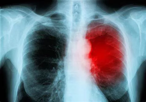
Chest X-rays are commonly used in clinical practice to evaluate heart and lung health. While they can provide valuable information, their utility in detecting myocarditis is somewhat limited. A chest X-ray can reveal certain findings that may suggest myocarditis, such as an enlarged heart (cardiomegaly), pulmonary congestion (fluid accumulation in the lungs), and signs of heart failure, such as pulmonary edema or pleural effusion (fluid around the lungs).
However, these findings are not specific to myocarditis and can occur in other cardiac and pulmonary conditions. For instance, cardiomegaly can result from conditions like dilated cardiomyopathy, while pulmonary congestion can be seen in congestive heart failure or pneumonia. Therefore, while a chest X-ray may raise suspicion for myocarditis, it cannot definitively confirm the diagnosis.
Limitations of Chest X-rays in Myocarditis Evaluation
Several factors contribute to the limitations of chest X-rays in evaluating myocarditis:
1. Lack of Specificity: The findings on a chest X-ray, such as cardiomegaly or pulmonary congestion, are nonspecific and can be caused by various cardiac and pulmonary conditions.
2. Inability to Visualize Myocardial Inflammation: Chest X-rays primarily focus on assessing the size and shape of the heart and detecting abnormalities in the lungs. They do not directly visualize inflammation or damage to the myocardium, which is the hallmark of myocarditis.
3. Need for Additional Imaging: In cases where myocarditis is suspected based on clinical symptoms and other tests, additional imaging modalities such as cardiac MRI (magnetic resonance imaging) or echocardiography are often necessary to assess myocardial function, detect inflammation, and rule out other cardiac abnormalities.
Complementary Diagnostic Tools for Myocarditis
While chest X-rays provide valuable information about cardiac and pulmonary anatomy, they are just one piece of the diagnostic puzzle for myocarditis. Healthcare providers may utilize several other tools and tests to evaluate patients with suspected myocarditis:
1. Electrocardiogram (ECG/EKG): This non-invasive test records the electrical activity of the heart and can detect abnormal rhythms (arrhythmias) or changes suggestive of myocardial damage.
2. Echocardiography (Echo): Also known as cardiac ultrasound, echocardiography uses sound waves to create images of the heart’s structure and function. It can assess heart chamber sizes, wall motion abnormalities, and the presence of fluid around the heart (pericardial effusion).
3. Cardiac MRI: Considered the gold standard for assessing myocardial inflammation and function, cardiac MRI provides detailed images of the heart muscle and can detect areas of inflammation (edema), scar tissue, and impaired contractility.
4. Laboratory Tests: Blood tests such as cardiac biomarkers (troponin, B-type natriuretic peptide) and inflammatory markers (C-reactive protein, erythrocyte sedimentation rate) can help assess heart muscle damage, inflammation levels, and overall cardiac function.
5. Endomyocardial Biopsy: In certain cases, a biopsy of the heart muscle (endomyocardial biopsy) may be performed to directly assess inflammation, identify the underlying cause (e.g., viral particles), and guide treatment decisions. However, this invasive procedure is typically reserved for severe or refractory cases of myocarditis.
Clinical Approach to Myocarditis Diagnosis
Given the complexities involved in diagnosing myocarditis, healthcare providers adopt a systematic approach to evaluate patients presenting with suspected myocarditis:
1. Clinical History and Physical Examination: A detailed history of symptoms, risk factors (e.g., recent viral illness, medication use), and a thorough physical examination are crucial initial steps in assessing myocarditis.
2. Diagnostic Tests: Based on clinical suspicion, healthcare providers may order diagnostic tests such as chest X-rays, electrocardiograms, echocardiography, cardiac MRI, and laboratory tests to evaluate cardiac function, detect inflammation, and identify potential causes.
3. Multidisciplinary Collaboration: Collaboration among cardiologists, infectious disease specialists, radiologists, and other healthcare professionals is essential for interpreting test results, determining the severity of myocarditis, identifying the underlying cause, and formulating an appropriate treatment plan.
4. Treatment and Monitoring: Treatment strategies for myocarditis depend on the underlying cause, severity of symptoms, and presence of complications such as heart failure or arrhythmias. Management may include supportive care, medications (such as anti-inflammatory drugs or immunosuppressants), lifestyle modifications, and close monitoring of cardiac function.
5. Patient Education and Follow-up: Educating patients about myocarditis, its potential complications, and the importance of adherence to treatment and follow-up appointments is crucial for optimal outcomes. Regular follow-up visits allow healthcare providers to monitor progress, adjust treatment as needed, and address any concerns or new symptoms.
Conclusion
While chest X-rays play a role in evaluating cardiac and pulmonary conditions, their ability to diagnose myocarditis is limited due to nonspecific findings and the inability to directly visualize myocardial inflammation. Healthcare providers employ a comprehensive approach to myocarditis diagnosis, utilizing a combination of clinical assessment, imaging studies (such as echocardiography and cardiac MRI), laboratory tests, and sometimes invasive procedures like endomyocardial biopsy. By integrating these diagnostic tools and collaborating across specialties, healthcare teams can accurately diagnose myocarditis, identify the underlying cause, and implement appropriate treatment strategies to improve patient outcomes and prevent complications.

