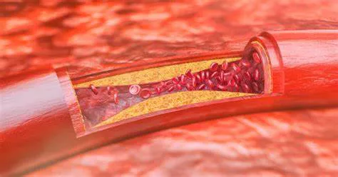
Coronary artery disease (CAD) is a condition where the coronary arteries that supply blood to the heart muscle become narrowed or blocked due to the buildup of plaque. This can lead to serious complications such as angina, heart attacks, and even sudden cardiac death. Early diagnosis and treatment are crucial in managing CAD and preventing its progression.
Doctors use a variety of tests and procedures to diagnose CAD, each tailored to the patient’s symptoms, risk factors, and overall health. This article will explore the methods doctors use to test for coronary artery disease in detail.
Symptoms And Risk Factors
Before delving into specific tests, it’s essential to understand the symptoms and risk factors that might prompt a doctor to investigate for CAD. Common symptoms include chest pain or discomfort (angina), shortness of breath, fatigue, and palpitations. Risk factors include high blood pressure, high cholesterol, smoking, diabetes, obesity, physical inactivity, and a family history of heart disease.
If a patient presents with these symptoms or risk factors, a doctor will likely recommend further testing to determine the presence of CAD.
Initial Evaluation And Physical Examination
The diagnostic process typically begins with a thorough medical history and physical examination. During the medical history, the doctor will ask about the patient’s symptoms, lifestyle, and family history of heart disease.
The physical examination may include listening to the heart and lungs, checking blood pressure, and looking for signs of other conditions that might contribute to heart disease.
Electrocardiogram (ECG or EKG)
An electrocardiogram (ECG or EKG) is one of the first tests performed when CAD is suspected. This test records the electrical activity of the heart and can reveal abnormalities such as arrhythmias, heart attacks, and other cardiac conditions.
While an ECG cannot definitively diagnose CAD, it can provide critical clues that further testing is needed.
Blood Tests
Blood tests are used to measure levels of cholesterol, triglycerides, blood sugar, and other markers that can indicate the presence of CAD. High levels of certain lipids, such as low-density lipoprotein (LDL) cholesterol, are associated with an increased risk of plaque buildup in the arteries. Blood tests can also detect markers of inflammation, such as C-reactive protein (CRP), which is linked to heart disease.
Chest X-ray
A chest X-ray can provide a picture of the heart, lungs, and blood vessels. While it’s not a specific test for CAD, it can help rule out other causes of symptoms such as lung conditions or an enlarged heart.
Non-invasive Diagnostic Tests
Stress Testing
Stress testing evaluates how the heart functions under physical stress. There are several types of stress tests:
Exercise Stress Test: The patient exercises on a treadmill or stationary bike while their heart rate, blood pressure, and ECG are monitored. This test can reveal how well the heart handles increased workload and can identify areas of reduced blood flow to the heart.
Nuclear Stress Test: This involves injecting a small amount of radioactive substance into the bloodstream. Images of the heart are taken at rest and after exercise to show areas with poor blood flow or damage.
Pharmacologic Stress Test: For patients unable to exercise, medication is given to simulate the effects of exercise on the heart. The heart’s response is then monitored using imaging techniques.
Echocardiogram
An echocardiogram uses ultrasound waves to create images of the heart. It provides detailed information about the heart’s structure and function, including the size and shape of the heart, the functioning of its valves, and the flow of blood through the chambers.
Stress echocardiography, a combination of an echocardiogram and a stress test, can show how well the heart pumps under stress.
CT Coronary Angiogram
A computed tomography (CT) coronary angiogram is a non-invasive imaging test that provides detailed pictures of the coronary arteries. It involves injecting a contrast dye into the bloodstream and then taking high-resolution images with a CT scanner.
This test can detect narrowed or blocked arteries and is highly effective in ruling out CAD in patients with a low to moderate risk.
Coronary Calcium Scan
Also known as a heart scan, this test uses a CT scanner to detect calcium deposits in the coronary arteries. The presence of calcium is a marker of atherosclerosis, a condition that can lead to CAD.
The amount of calcium is measured to produce a score that reflects the risk of CAD.
Invasive Diagnostic Tests
Coronary Angiography
Coronary angiography is considered the gold standard for diagnosing CAD. This invasive procedure involves threading a catheter through the blood vessels to the coronary arteries. A contrast dye is then injected, and X-ray images are taken to visualize the arteries. This test provides detailed information about the location and severity of any blockages and can also guide treatment decisions such as angioplasty or stenting.
Intravascular Ultrasound (IVUS)
Intravascular ultrasound (IVUS) is often used in conjunction with coronary angiography. It involves inserting a tiny ultrasound probe into the coronary arteries via a catheter. IVUS provides detailed images of the artery walls and can help assess the extent of plaque buildup and the structure of the arteries.
Fractional Flow Reserve (FFR)
Fractional flow reserve (FFR) is a technique used during coronary angiography to measure blood pressure differences across a coronary artery stenosis. By calculating the FFR, doctors can determine the functional significance of the stenosis and decide whether it needs to be treated with angioplasty or stenting.
Emerging Diagnostic Techniques
Magnetic Resonance Imaging (MRI)
Cardiac magnetic resonance imaging (MRI) is a non-invasive test that provides detailed images of the heart and blood vessels without the use of radiation. It can assess the structure and function of the heart, detect areas of damage, and evaluate blood flow through the coronary arteries.
While not yet as widely used as other tests, MRI is becoming an important tool in the diagnosis and management of CAD.
Optical Coherence Tomography (OCT)
Optical coherence tomography (OCT) is a high-resolution imaging technique that can visualize the inside of coronary arteries.
It uses light waves to create detailed cross-sectional images and is often used during angiography to provide additional information about plaque characteristics and vessel structure.
Conclusion
Diagnosing coronary artery disease involves a combination of patient history, physical examination, and a variety of diagnostic tests. The choice of tests depends on the patient’s symptoms, risk factors, and overall health. From non-invasive methods like stress testing and CT angiography to invasive procedures like coronary angiography and IVUS, each test provides valuable information that helps doctors assess the presence and severity of CAD.

