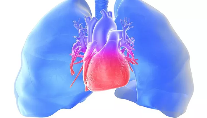
Scleroderma, also known as systemic sclerosis, is a complex autoimmune disease characterized by the hardening and tightening of the skin and connective tissues. This condition affects not only the skin but also various internal organs, leading to significant morbidity and mortality. One of the most serious complications associated with scleroderma is pulmonary hypertension (PH). Pulmonary hypertension is a type of high blood pressure that affects the arteries in the lungs and the right side of the heart. Understanding the mechanisms by which scleroderma leads to pulmonary hypertension is crucial for the development of effective treatments and management strategies.
The Pathophysiology of Scleroderma
Scleroderma is an autoimmune disorder in which the immune system mistakenly attacks the body’s own tissues. This leads to the overproduction of collagen, a protein that makes up the connective tissues, causing the skin and other organs to harden and thicken. The pathophysiology of scleroderma involves a combination of vascular abnormalities, immune system dysregulation, and fibrosis.
Vascular Abnormalities
One of the hallmarks of scleroderma is damage to the small blood vessels, known as microvascular injury. This damage is thought to be an early event in the disease process and plays a central role in the development of pulmonary hypertension.
The endothelial cells lining the blood vessels are particularly affected, leading to:
Endothelial Dysfunction: The endothelial cells become dysfunctional, reducing their ability to regulate vascular tone and maintain a barrier against harmful substances.
Vasculopathy: This refers to the pathological changes in the blood vessels, including narrowing, reduced elasticity, and the formation of fibrous tissue.
see also: How Does Left Heart Failure Cause Pulmonary Hypertension
Immune System Dysregulation
In scleroderma, the immune system produces autoantibodies that attack the body’s own tissues. This autoimmune response leads to chronic inflammation and tissue damage. The release of pro-inflammatory cytokines and growth factors contributes to the ongoing vascular damage and fibrosis.
Fibrosis
Fibrosis is the excessive formation of connective tissue, leading to the hardening and thickening of the affected tissues. In scleroderma, fibrosis occurs not only in the skin but also in various internal organs, including the lungs. The fibrotic process is driven by the overproduction of collagen and other extracellular matrix components, which replace the normal tissue architecture.
Mechanisms of Pulmonary Hypertension in Scleroderma
Pulmonary hypertension in scleroderma results from a combination of vascular, immune, and fibrotic processes. The following mechanisms are involved:
Vascular Remodeling
The structural changes in the pulmonary blood vessels, known as vascular remodeling, are central to the development of pulmonary hypertension. These changes include:
Intimal Proliferation: The innermost layer of the blood vessels (intima) thickens due to the proliferation of smooth muscle cells and fibroblasts.
Medial Hypertrophy: The middle layer of the blood vessels (media) becomes thicker as smooth muscle cells proliferate and accumulate.
Adventitial Fibrosis: The outer layer of the blood vessels (adventitia) undergoes fibrosis, further contributing to the narrowing of the vessel lumen.
Endothelial Dysfunction
Endothelial dysfunction in scleroderma leads to:
Reduced Nitric Oxide Production: Nitric oxide is a vasodilator that helps maintain the patency of blood vessels. Reduced production of nitric oxide results in vasoconstriction.
Increased Endothelin-1 Production: Endothelin-1 is a potent vasoconstrictor that promotes the narrowing of blood vessels and stimulates smooth muscle cell proliferation.
Inflammation and Autoimmunity
Chronic inflammation and the presence of autoantibodies in scleroderma contribute to vascular injury and remodeling.
Pro-inflammatory cytokines, such as interleukin-6 (IL-6) and tumor necrosis factor-alpha (TNF-alpha), play a significant role in promoting inflammation and vascular changes.
Hypoxia
Hypoxia, or low oxygen levels in the tissues, can also contribute to the development of pulmonary hypertension. In scleroderma, hypoxia results from impaired lung function due to interstitial lung disease (ILD) and fibrosis. The body responds to hypoxia by constricting the pulmonary arteries, which increases pulmonary vascular resistance and contributes to pulmonary hypertension.
Clinical Presentation of Pulmonary Hypertension in Scleroderma
Pulmonary hypertension in scleroderma patients can present with a variety of symptoms, which are often non-specific and can overlap with other manifestations of the disease. Common symptoms include:
Dyspnea: Shortness of breath, especially during exertion, is a common symptom of pulmonary hypertension.
Fatigue: Patients often experience severe fatigue, which can be debilitating.
Chest Pain: Chest pain or discomfort may occur due to increased pressure in the pulmonary arteries.
Syncope: Fainting or lightheadedness can occur due to reduced blood flow to the brain.
Edema: Swelling in the legs and ankles can result from right heart failure, a complication of pulmonary hypertension.
Diagnostic Evaluation
The diagnosis of pulmonary hypertension in scleroderma involves a combination of clinical evaluation, imaging studies, and hemodynamic assessment. Key diagnostic tools include:
Echocardiography: This non-invasive imaging technique uses ultrasound to evaluate the structure and function of the heart and estimate the pulmonary artery pressure.
Right Heart Catheterization: This invasive procedure is considered the gold standard for diagnosing pulmonary hypertension. It measures the pressure in the pulmonary arteries and assesses the hemodynamic parameters.
Pulmonary Function Tests: These tests evaluate lung function and can help identify the presence of interstitial lung disease, which is common in scleroderma patients.
High-Resolution CT Scan: A high-resolution computed tomography (CT) scan of the chest can identify interstitial lung disease and other lung abnormalities.
Management of Pulmonary Hypertension in Scleroderma
The management of pulmonary hypertension in scleroderma is challenging and requires a multidisciplinary approach.
Treatment strategies aim to improve symptoms, enhance quality of life, and slow disease progression. Key components of management include:
Medications
Several classes of medications are used to treat pulmonary hypertension, including:
Endothelin Receptor Antagonists (ERAs): These drugs, such as bosentan and ambrisentan, block the effects of endothelin-1, reducing vasoconstriction and vascular remodeling.
Phosphodiesterase-5 Inhibitors (PDE-5 inhibitors): Medications like sildenafil and tadalafil enhance the effects of nitric oxide, promoting vasodilation and reducing pulmonary artery pressure.
Prostacyclin Analogues: These drugs, including epoprostenol and treprostinil, mimic the effects of prostacyclin, a natural vasodilator, and inhibit platelet aggregation.
Soluble Guanylate Cyclase Stimulators: Riociguat is a drug that enhances the effects of nitric oxide and promotes vasodilation.
Oxygen Therapy
For patients with significant hypoxia, supplemental oxygen therapy may be necessary to maintain adequate oxygen levels and reduce pulmonary artery pressure.
Immunosuppressive Therapy
In cases where inflammation plays a significant role, immunosuppressive medications, such as corticosteroids and mycophenolate mofetil, may be used to reduce immune system activity and inflammation.
Lifestyle Modifications
Lifestyle modifications can help manage symptoms and improve overall health. Recommendations include:
Regular Exercise: Tailored exercise programs can help improve cardiovascular fitness and reduce symptoms of fatigue.
Healthy Diet: A balanced diet can support overall health and help manage comorbid conditions.
Smoking Cessation: Smoking can exacerbate pulmonary hypertension and other complications of scleroderma.
Monitoring and Follow-up
Regular follow-up with a multidisciplinary team, including rheumatologists, pulmonologists, and cardiologists, is essential for monitoring disease progression and adjusting treatment as needed.
Conclusion
Pulmonary hypertension is a serious and potentially life-threatening complication of scleroderma. The development of pulmonary hypertension in scleroderma involves complex interactions between vascular abnormalities, immune system dysregulation, and fibrosis. Early diagnosis and comprehensive management are crucial to improving outcomes and quality of life for patients with scleroderma-associated pulmonary hypertension. Ongoing research is essential to better understand the underlying mechanisms and to develop more effective treatments for this challenging condition.


