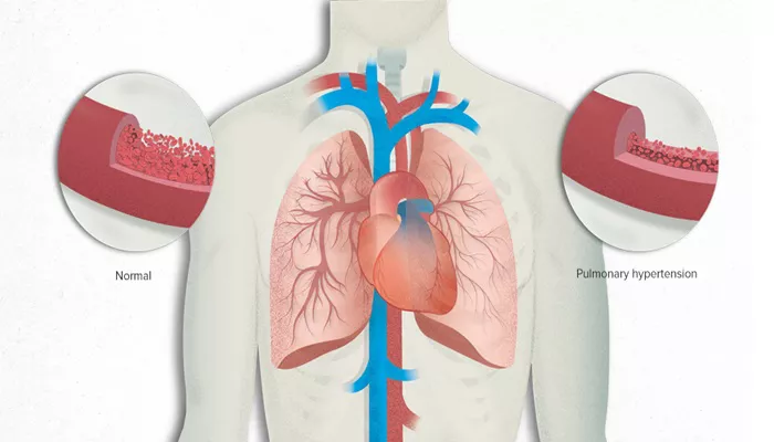
Pulmonary hypertension (PH) is a serious and potentially life-threatening condition characterized by elevated blood pressure in the arteries of the lungs. Early diagnosis is crucial for effective management and treatment. Testing for pulmonary hypertension involves a combination of clinical evaluation, imaging studies, and specialized tests. This article will detail the various methods and procedures used to diagnose pulmonary hypertension, including their purposes, processes, and interpretations.
Understanding Pulmonary Hypertension
Before diving into the diagnostic methods, it is essential to understand what pulmonary hypertension is and why it is a concern. Pulmonary hypertension occurs when the blood vessels in the lungs become narrowed, blocked, or damaged, leading to increased resistance to blood flow. This condition places additional strain on the right side of the heart, which can result in heart failure if left untreated.
How Do You Test for Pulmonary Hypertension?
1. Clinical Evaluation
Medical History and Physical Examination
The diagnostic process begins with a thorough medical history and physical examination. The physician will inquire about the patient’s symptoms, family history, and any pre-existing conditions that might contribute to pulmonary hypertension, such as chronic obstructive pulmonary disease (COPD), heart disease, or connective tissue disorders.
During the physical examination, the doctor will look for signs that might indicate pulmonary hypertension, including:
Jugular venous distention: Swelling of the neck veins.
Hepatomegaly: Enlargement of the liver.
Peripheral edema: Swelling in the legs and ankles.
Cyanosis: Bluish discoloration of the skin due to low oxygen levels.
SEE ALSO: How Does Hypertension Cause Proteinuria?
2. Non-Invasive Tests
Electrocardiogram (ECG)
An electrocardiogram is a simple and quick test that records the electrical activity of the heart. It helps identify abnormalities in heart rhythm and structure. In pulmonary hypertension, the ECG may show:
Right ventricular hypertrophy (enlargement of the right side of the heart).
Right axis deviation.
Signs of right atrial enlargement.
Chest X-ray
A chest X-ray provides an image of the heart, lungs, and blood vessels.
It can help identify changes in the size and shape of the heart and pulmonary arteries. In cases of pulmonary hypertension, a chest X-ray might reveal:
Enlargement of the right ventricle and pulmonary arteries.
Signs of underlying lung disease.
Echocardiogram
An echocardiogram, or ultrasound of the heart, is a crucial non-invasive test for diagnosing pulmonary hypertension. It uses sound waves to create detailed images of the heart’s structure and function. The echocardiogram can estimate the pressure in the pulmonary arteries and assess the right ventricular function. Key findings that suggest pulmonary hypertension include:
Elevated right ventricular systolic pressure (RVSP).
Enlargement of the right ventricle and right atrium.
Abnormal movement of the interventricular septum.
Pulmonary Function Tests (PFTs)
Pulmonary function tests measure how well the lungs are working.
These tests include spirometry, which assesses the amount and speed of air a person can exhale, and diffusion capacity, which measures how well oxygen passes from the lungs into the blood. PFTs can help identify underlying lung diseases contributing to pulmonary hypertension.
Six-Minute Walk Test (6MWT)
The six-minute walk test evaluates a patient’s exercise capacity and endurance. During this test, the patient walks for six minutes, and the distance covered is recorded. The test helps determine the severity of pulmonary hypertension and monitors the effectiveness of treatment.
3. Advanced Imaging Studies
Computed Tomography (CT) Scan
A computed tomography scan provides detailed cross-sectional images of the chest, including the lungs and blood vessels.
A high-resolution CT scan can detect underlying lung conditions such as interstitial lung disease or pulmonary embolism, which may contribute to pulmonary hypertension.
Ventilation-Perfusion (V/Q) Scan
A ventilation-perfusion scan is a nuclear medicine test that evaluates the airflow (ventilation) and blood flow (perfusion) in the lungs. This test is particularly useful in diagnosing chronic thromboembolic pulmonary hypertension (CTEPH), a form of PH caused by blood clots in the pulmonary arteries. The V/Q scan can identify areas of the lungs that are receiving adequate ventilation but have impaired blood flow due to clots.
Magnetic Resonance Imaging (MRI)
Cardiac magnetic resonance imaging provides detailed images of the heart and pulmonary arteries without using radiation.
It can assess right ventricular function, measure blood flow, and evaluate the size and shape of the heart chambers. MRI is particularly useful for patients who cannot undergo other imaging tests due to allergies or kidney problems.
4. Invasive Tests
Right Heart Catheterization (RHC)
Right heart catheterization is considered the gold standard for diagnosing pulmonary hypertension. This invasive procedure measures the pressures in the right side of the heart and pulmonary arteries directly. During RHC, a thin, flexible tube (catheter) is inserted through a vein in the neck, arm, or groin and guided into the right side of the heart and pulmonary arteries. The following parameters are measured:
Pulmonary artery pressure (PAP).
Pulmonary capillary wedge pressure (PCWP).
Cardiac output (CO).
Pulmonary vascular resistance (PVR).
Right heart catheterization not only confirms the diagnosis of pulmonary hypertension but also helps determine its severity and guide treatment decisions.
Acute Vasoreactivity Testing
During right heart catheterization, acute vasoreactivity testing may be performed to assess the responsiveness of the pulmonary arteries to vasodilator medications. This test helps identify patients with pulmonary arterial hypertension (PAH) who may benefit from calcium channel blockers. In this test, a vasodilator such as inhaled nitric oxide, intravenous epoprostenol, or intravenous adenosine is administered, and the changes in pulmonary artery pressure and cardiac output are measured.
Blood Tests
Blood tests are often conducted to rule out other conditions that may cause or contribute to pulmonary hypertension.
These tests can include:
Complete blood count (CBC): To check for anemia or polycythemia.
Liver function tests: To evaluate liver health.
Thyroid function tests: To assess thyroid hormone levels.
Autoimmune markers: To detect connective tissue diseases such as scleroderma or lupus.
B-type natriuretic peptide (BNP): Elevated levels of BNP can indicate heart failure, which is common in advanced pulmonary hypertension.
Genetic Testing
In some cases, genetic testing may be recommended, especially if there is a family history of pulmonary arterial hypertension (PAH).
Mutations in specific genes, such as BMPR2, can predispose individuals to PAH. Identifying these mutations can help in understanding the cause of the disease and guiding family counseling and management.
Conclusion
Testing for pulmonary hypertension involves a comprehensive approach that includes clinical evaluation, non-invasive tests, advanced imaging studies, and invasive procedures. Early and accurate diagnosis is crucial for effective management and improving the quality of life for patients with pulmonary hypertension. Each diagnostic method plays a vital role in understanding the severity and underlying causes of the condition, guiding appropriate treatment strategies.

