The carotid arteries are the main blood vessels responsible for supplying oxygenated blood to the brain. These arteries run along the sides of the neck and can become narrowed or blocked due to the buildup of fatty deposits called plaque. This condition, known as carotid artery disease or carotid stenosis, can lead to a reduced blood flow to the brain and increase the risk of stroke. Understanding the normal levels of carotid artery blockage is crucial for early detection and prevention of this potentially life-threatening condition.
Anatomy of The Carotid Arteries
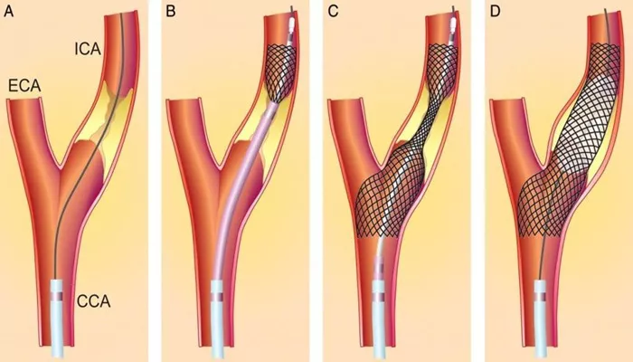
The carotid arteries are divided into two main branches: the common carotid artery and the internal carotid artery. The common carotid artery runs along the front of the neck and splits into the internal and external carotid arteries at the level of the fourth cervical vertebra. The internal carotid artery supplies blood to the brain, while the external carotid artery supplies blood to the face and scalp.
Causes of Carotid Artery Blockage
Carotid artery blockage is primarily caused by atherosclerosis, a condition in which plaque builds up inside the arteries. Plaque is made up of cholesterol, fatty substances, cellular waste products, calcium, and fibrin. Over time, this buildup narrows and hardens the arteries, reducing blood flow to the brain.
Other risk factors for carotid artery disease include:
- High blood pressure
- High cholesterol
- Diabetes
- Smoking
- Obesity
- Family history of carotid artery disease or stroke
- Increasing age
Measuring Carotid Artery Blockage
Carotid artery blockage is typically measured using a percentage scale, with 0% indicating no blockage and 100% indicating a complete blockage or occlusion. The degree of blockage is determined by comparing the narrowest part of the artery to a normal, healthy section of the artery.
Several imaging techniques can be used to assess carotid artery blockage, including:
Carotid ultrasound: This non-invasive test uses sound waves to create images of the carotid arteries and measure the degree of blockage.
Computed tomography angiography (CTA): This test uses X-rays and a contrast dye to create detailed images of the carotid arteries.
SEE ALSO: What Is Percutaneous Coronary Intervention?
Magnetic resonance angiography (MRA): This test uses magnetic fields and radio waves to create images of the carotid arteries.
Cerebral angiography: This invasive test involves inserting a catheter into a blood vessel in the groin or arm and threading it up to the carotid arteries. A contrast dye is then injected, and X-rays are taken to create detailed images of the arteries.
Normal Levels of Carotid Artery Blockage
In general, a carotid artery blockage of less than 50% is considered normal and does not typically require treatment.
However, it’s important to note that even a small amount of plaque buildup can increase the risk of stroke over time.
The following guidelines are commonly used to classify the degree of carotid artery blockage:
Less than 50% blockage: Normal or mild blockage
50% to 69% blockage: Moderate blockage
70% to 99% blockage: Severe blockage
100% blockage: Complete blockage or occlusion
It’s important to note that these guidelines are not absolute, and individual risk factors and symptoms should be taken into account when determining the appropriate course of treatment.
Symptoms of Carotid Artery Blockage
In many cases, carotid artery blockage does not cause any symptoms until it becomes severe. However, some individuals may experience symptoms such as:
Sudden weakness or numbness in the face, arm, or leg, often on one side of the body
Difficulty speaking or understanding speech
Sudden trouble seeing in one or both eyes
Sudden severe headache with no known cause
Difficulty swallowing
Loss of balance or coordination
These symptoms may indicate a transient ischemic attack (TIA) or a stroke. It’s crucial to seek immediate medical attention if these symptoms occur, as prompt treatment can help prevent permanent brain damage and reduce the risk of a more severe stroke.
Treatment Options for Carotid Artery Blockage
Treatment for carotid artery blockage depends on the severity of the condition and the individual’s risk factors. In cases of mild to moderate blockage, treatment may focus on lifestyle changes and medication to manage risk factors and prevent further plaque buildup. These measures may include:
Quitting smoking
Eating a healthy diet low in saturated fat and cholesterol
Exercising regularly
Maintaining a healthy weight
Taking medications to control blood pressure, cholesterol, and diabetes
In cases of severe blockage (70% or greater), treatment may involve surgical procedures to remove the plaque and restore blood flow to the brain. These procedures include:
Carotid endarterectomy: This surgery involves making an incision in the neck and removing the plaque from the carotid artery.
Carotid artery stenting: This minimally invasive procedure involves inserting a small mesh tube called a stent into the carotid artery to keep it open and improve blood flow to the brain.
The decision to undergo surgery depends on several factors, including the individual’s age, overall health, and the location and severity of the blockage. In some cases, the risks of surgery may outweigh the potential benefits, and medical management may be the preferred course of treatment.
Preventing Carotid Artery Blockage
The best way to prevent carotid artery blockage is to maintain a healthy lifestyle and manage risk factors. This may include:
Quitting smoking
Eating a healthy diet low in saturated fat and cholesterol
Exercising regularly
Maintaining a healthy weight
Managing blood pressure, cholesterol, and diabetes
Regular checkups with a healthcare provider can also help detect carotid artery disease early and prevent complications such as stroke.
Conclusion
Carotid artery blockage is a common condition that can lead to serious complications, including stroke. Understanding the normal levels of carotid artery blockage is crucial for early detection and prevention of this potentially life-threatening condition. While a blockage of less than 50% is considered normal, even a small amount of plaque buildup can increase the risk of stroke over time.

