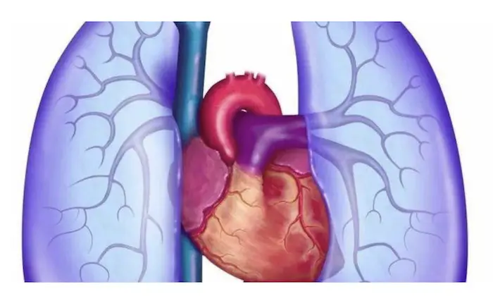
Atrial septal defect (ASD) is a congenital heart defect characterized by an opening in the atrial septum, the wall that separates the two upper chambers of the heart. This defect allows blood to flow from the left atrium to the right atrium, leading to increased blood flow to the right side of the heart and the lungs. One of the significant complications associated with ASD is the development of pulmonary hypertension (PH), a condition characterized by elevated blood pressure in the pulmonary arteries. This article explores the relationship between ASD and pulmonary hypertension, including the mechanisms by which ASD can lead to PH, the symptoms, diagnosis, treatment options, and the long-term implications for patient health.
Understanding Atrial Septal Defect (ASD)
Definition
An atrial septal defect is a congenital heart defect that results in an abnormal opening in the atrial septum. This defect can vary in size and may be classified into several types, including.
Ostium Secundum ASD: The most common type, located in the middle of the septum.
Ostium Primum ASD: Often associated with other congenital heart defects, located lower in the septum.
Sinus Venosus ASD: Located near the entrance of the superior vena cava, often associated with partial anomalous pulmonary venous return.
Common Atrium: A rare and complex form where there is no septum between the atria.
Causes
The exact cause of ASD is often unknown, but several factors may contribute to its development, including:
Genetic Factors: Family history of congenital heart defects may increase the risk.
Environmental Factors: Maternal exposure to certain medications, alcohol, or infections during pregnancy may be associated with an increased risk of ASD.
Symptoms
Many individuals with ASD remain asymptomatic for years, especially if the defect is small. However, larger ASDs can lead to symptoms such as.
Shortness of Breath: Particularly during exertion or physical activity.
Fatigue: A general feeling of tiredness and decreased exercise tolerance.
Palpitations: Irregular heartbeats or sensations of rapid heart rate.
Frequent Respiratory Infections: Due to increased blood flow to the lungs.
Swelling: In the legs, abdomen, or veins in the neck.
Understanding Pulmonary Hypertension (PH)
Definition
Pulmonary hypertension is defined as elevated blood pressure in the pulmonary arteries, typically characterized by a mean pulmonary arterial pressure (mPAP) greater than 25 mmHg at rest, as measured by right heart catheterization. Normal pulmonary arterial pressure ranges from 8 to 20 mmHg.
Causes
Pulmonary hypertension can be classified into five groups based on the underlying causes:
Group 1: Pulmonary Arterial Hypertension (PAH): Includes idiopathic and heritable forms, as well as those associated with connective tissue diseases, congenital heart diseases, and other conditions.
Group 2: Pulmonary Hypertension due to Left Heart Disease: Results from left-sided heart conditions.
Group 3: Pulmonary Hypertension due to Lung Diseases and/or Hypoxia: Associated with chronic lung diseases and low oxygen levels.
Group 4: Chronic Thromboembolic Pulmonary Hypertension (CTEPH): Results from blood clots in the pulmonary arteries.
Group 5: Pulmonary Hypertension with Unclear Multifactorial Mechanisms: Includes various other conditions that do not fit into the above categories.
Symptoms
Symptoms of pulmonary hypertension may include:
Shortness of Breath: Especially during physical activity.
Fatigue: A persistent feeling of tiredness.
Chest Pain: Discomfort or pain in the chest.
Palpitations: Awareness of an irregular or rapid heartbeat.
Swelling: In the ankles, legs, or abdomen due to fluid retention.
Cyanosis: A bluish tint to the lips and skin, indicating inadequate oxygenation.
The Link Between Atrial Septal Defect and Pulmonary Hypertension
Mechanisms Linking ASD to Pulmonary Hypertension
Increased Right Heart Volume Load: The left-to-right shunt caused by ASD results in increased blood flow to the right atrium and right ventricle. Over time, this increased volume load can lead to right ventricular dilation and hypertrophy.
Increased Pulmonary Blood Flow: The excess blood flow to the pulmonary circulation can lead to increased pressure in the pulmonary arteries. This increased pressure can result in pulmonary vascular remodeling and increased resistance to blood flow.
Pulmonary Vascular Remodeling: The chronic increase in blood flow and pressure can lead to structural changes in the pulmonary vasculature, including endothelial dysfunction, smooth muscle proliferation, and fibrosis. These changes contribute to increased pulmonary vascular resistance and the development of pulmonary hypertension.
Hypoxia and Inflammation: Increased blood flow to the lungs can lead to hypoxia and inflammation, further exacerbating pulmonary vascular changes and contributing to the development of pulmonary hypertension.
Clinical Evidence
Research has shown that individuals with untreated ASD are at a significantly increased risk of developing pulmonary hypertension. Key findings include.
Prevalence of PH in ASD Patients: Studies have demonstrated that up to 30% of patients with ASD may develop pulmonary hypertension, especially if the defect is large and left untreated.
Severity Correlation: The severity of pulmonary hypertension often correlates with the size of the ASD and the volume of left-to-right shunt.
Reversibility: In some cases, pulmonary hypertension may be reversible after closure of the ASD, particularly if it is done before significant irreversible changes occur in the pulmonary vasculature.
Diagnosis of Pulmonary Hypertension in the Context of ASD
Clinical Evaluation
Diagnosing pulmonary hypertension in patients with ASD involves a comprehensive clinical evaluation, including:
Medical History: Gathering information on symptoms, family history, and risk factors for congenital heart disease and pulmonary hypertension.
Physical Examination: Checking vital signs, heart sounds, and signs of heart failure, such as peripheral edema or elevated jugular venous pressure.
Diagnostic Tests
Echocardiography: This non-invasive imaging test is often the first step in diagnosing ASD and assessing right heart function. It can estimate pulmonary artery pressure and visualize the shunt.
Right Heart Catheterization: This invasive procedure is considered the gold standard for diagnosing pulmonary hypertension. It directly measures pressures in the right atrium, right ventricle, pulmonary artery, and pulmonary capillary wedge pressure.
Chest X-ray: This imaging study can reveal signs of right heart enlargement and increased pulmonary vascular markings.
Pulmonary Function Tests: These tests assess lung function and can help rule out other causes of dyspnea.
Blood Tests: Assessing levels of natriuretic peptides (e.g., BNP or NT-proBNP) can help evaluate heart failure and its severity.
Treatment of Atrial Septal Defect and Pulmonary Hypertension
Treatment of Atrial Septal Defect
Observation: In small ASDs, particularly in asymptomatic patients, careful monitoring may be sufficient.
Percutaneous Closure: For moderate to large ASDs, a minimally invasive procedure using a catheter to place a closure device may be performed.
Surgical Repair: In cases where percutaneous closure is not feasible or in patients with significant symptoms, surgical repair may be necessary.
Management of Pulmonary Hypertension
Pharmacological Treatments: If pulmonary hypertension persists after ASD closure, medications may be necessary to manage blood pressure and improve symptoms. Common classes of medications include.
Endothelin Receptor Antagonists (ERAs): Help dilate blood vessels and reduce pulmonary artery pressure.
Phosphodiesterase-5 Inhibitors (PDE-5 Inhibitors): Enhance the effects of nitric oxide, leading to vasodilation.
Prostacyclin Analogues: Mimic the effects of prostacyclin, a natural vasodilator.
Oxygen Therapy: Supplemental oxygen may be necessary for patients with low oxygen saturation levels.
Lifestyle Modifications: Patients should be encouraged to engage in regular exercise, follow a heart-healthy diet, and avoid smoking.
Monitoring and Follow-Up: Regular follow-up appointments are essential for monitoring pulmonary pressures and adjusting treatment plans as necessary.
Implications for Patient Health
Quality of Life
The interplay between atrial septal defect and pulmonary hypertension can significantly impact a patient’s quality of life. Symptoms such as shortness of breath, fatigue, and reduced exercise tolerance can limit daily activities and social interactions. Effective management of both conditions is essential to improve overall well-being.
Long-Term Health Consequences
Untreated ASD and pulmonary hypertension can lead to serious long-term health consequences, including:
Increased Risk of Heart Failure: The volume overload on the right heart can lead to right ventricular failure, particularly in older patients or those with other comorbidities.
Increased Risk of Arrhythmias: Right atrial enlargement and structural changes can predispose patients to atrial fibrillation and other arrhythmias.
Cognitive Impairment: Chronic hypoxia and reduced cardiac output can contribute to cognitive decline and other neurological issues.
Metabolic Syndrome: The combination of ASD and pulmonary hypertension may contribute to the development of metabolic syndrome, increasing the risk of diabetes and other metabolic disorders.
Research and Future Directions
Ongoing research is essential to further elucidate the complex relationship between atrial septal defect and pulmonary hypertension. Areas of focus may include.
Mechanistic Studies: Investigating the underlying biological mechanisms linking ASD to pulmonary hypertension and cardiovascular disease.
Longitudinal Studies: Assessing the long-term cardiovascular outcomes of patients with ASD and pulmonary hypertension to better understand the trajectory of these conditions.
Intervention Studies: Evaluating the effectiveness of various treatment modalities for ASD and their impact on pulmonary pressure control and overall cardiovascular health.
Conclusion
Atrial septal defect (ASD) is a congenital heart defect that can lead to significant complications, including pulmonary hypertension. The mechanisms linking ASD to pulmonary hypertension involve increased right heart volume load, increased pulmonary blood flow, vascular remodeling, and systemic inflammation.
Recognizing the relationship between these two conditions is crucial for healthcare providers in diagnosing and managing affected patients. Through a combination of effective ASD treatment, lifestyle modifications, and careful management of pulmonary hypertension, healthcare providers can help mitigate the effects of both conditions and improve patient outcomes.
As our understanding of atrial septal defect and its cardiovascular implications continues to evolve, it is vital to prioritize research, education, and awareness to enhance the management of patients suffering from these interconnected health issues. By doing so, we can work towards better health outcomes and improved quality of life for individuals affected by atrial septal defect and pulmonary hypertension.
Related Topics:


