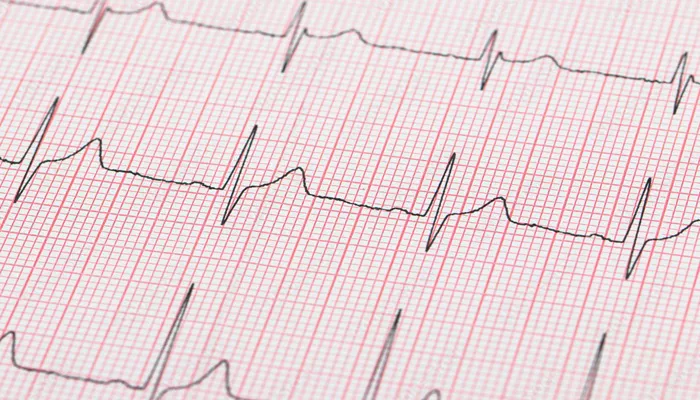
An Electrocardiogram (ECG or EKG) is a medical test that measures the electrical activity of the heart over a period of time. It is one of the most commonly performed diagnostic procedures in cardiology, providing valuable information about the heart’s rhythm, electrical impulses, and overall health. The ECG records the heart’s electrical signals through electrodes placed on the skin, creating a graphical representation of the heart’s activity. This non-invasive test is a crucial tool for diagnosing various heart conditions, including arrhythmias, heart attacks, and other cardiovascular disorders.
In this article, we will explore in detail what an Electrocardiogram is, how it works, its clinical significance, the procedure involved, and how to interpret the results.
How Does An Electrocardiogram Work?
An Electrocardiogram works by detecting the electrical impulses that trigger each heartbeat. These impulses are generated by the heart’s natural pacemaker, the sinoatrial (SA) node, which sends electrical signals through the heart muscle. As these electrical impulses pass through different parts of the heart, they cause the heart muscles to contract and pump blood.
The ECG test uses electrodes attached to the skin to detect and record the electrical activity. These electrodes are typically placed on specific areas of the body: the chest, arms, and legs. The electrical signals from the heart are transmitted through these electrodes to an ECG machine, which then produces a visual record of the signals in the form of a waveform.
Why Is An Electrocardiogram Important?
An ECG is an essential diagnostic tool that provides a wealth of information about the heart’s electrical activity and overall health. By analyzing the waveform generated by the ECG, a healthcare provider can detect a variety of heart conditions, such as:
Arrhythmias: Abnormal heart rhythms, such as atrial fibrillation, ventricular tachycardia, and other irregular heartbeats, can be identified through changes in the ECG waveform.
Heart Attacks (Myocardial Infarction): An ECG can help diagnose a heart attack by detecting changes in the electrical patterns caused by damage to the heart muscle.
Electrolyte Imbalances: Abnormalities in the ECG can sometimes indicate issues with electrolyte levels in the blood, such as potassium or calcium imbalances.
Heart Enlargement: The ECG can also show signs of an enlarged heart (hypertrophy), which may be a sign of heart disease or high blood pressure.
Pericarditis: Inflammation of the pericardium (the lining around the heart) can alter the electrical activity of the heart, and this can be seen in an ECG.
Long QT Syndrome: A condition that can increase the risk of sudden cardiac arrest can also be diagnosed by changes in the QT interval, which is visible on an ECG.
Types of Electrocardiograms
There are different types of ECGs, each suited for different purposes:
1. Resting ECG
A standard ECG performed while the patient is lying down and at rest. This is the most common type and is usually done in a clinic or hospital setting.
2. Stress (Exercise) ECG
A stress test, also called an exercise ECG, is performed while the patient exercises on a treadmill or stationary bike. This test helps evaluate how the heart responds to physical stress and is often used to detect coronary artery disease.
3. Holter Monitor
A Holter monitor is a portable ECG device that records the heart’s activity continuously for 24 to 48 hours or longer. It is often used to monitor heart rhythm over an extended period, particularly in cases of intermittent arrhythmias.
4. Event Recorder
An event recorder is similar to a Holter monitor but is worn for longer periods, sometimes weeks. It is used for patients who experience symptoms infrequently and helps detect abnormal heart rhythms during the event.
The Components of an ECG Waveform
The waveform produced by an ECG consists of several distinct components, each representing a phase of the heart’s electrical cycle:
P Wave: This represents the depolarization (or activation) of the atria, the upper chambers of the heart.
QRS Complex: The QRS complex represents the depolarization of the ventricles, the lower chambers of the heart, which is the most prominent and important part of the ECG. It is also associated with the contraction of the heart.
T Wave: This represents the repolarization (or recovery) of the ventricles, where the heart muscles reset in preparation for the next heartbeat.
U Wave (in some cases): This is sometimes seen following the T wave, although its exact origin and significance are not always clear.
Together, these components form a complete cycle that represents the electrical activity of a single heartbeat.
Early Detection And Monitoring
An ECG is useful not only for diagnosing existing heart conditions but also for detecting heart problems before symptoms appear. For example, a routine ECG might reveal signs of a heart condition in someone who feels healthy, allowing for early intervention and prevention of more severe issues down the road.
Additionally, the ECG is often used to monitor patients with existing heart conditions to track their heart’s health over time, assess the effectiveness of treatments, and detect any new or emerging issues.
The ECG Procedure: What to Expect
The ECG test is simple, fast, and non-invasive. Here’s what you can expect during the procedure:
1. Preparation
Before the test begins, the technician or nurse will explain the procedure and ask you to remove any clothing that covers your chest, arms, and legs. You will be provided with a gown to wear during the test.
2. Electrode Placement
Once you’re ready, the healthcare provider will attach small adhesive electrodes to your skin at specific locations on your chest, arms, and legs. The electrodes are designed to detect the electrical impulses from your heart.
Chest Electrodes: Typically, 6 electrodes are placed on your chest at various points to get a close-up reading of your heart’s electrical activity.
Limbs Electrodes: Four electrodes are placed on your limbs (two on each arm and leg) to capture the electrical signals from a different angle.
3. Recording the ECG
Once the electrodes are in place, the ECG machine will record your heart’s electrical activity. The test itself typically takes only about 5 to 10 minutes. You may be asked to lie still and breathe normally during this time to ensure the most accurate results.
4. After the Test
After the test, the electrodes are removed, and you are free to go about your normal activities. The test is completely painless, and there is no recovery time required.
Interpreting the Electrocardiogram
Once the ECG is recorded, it is analyzed by a healthcare provider, usually a cardiologist, who will look for various abnormalities in the waveform. The doctor will check for:
Heart rate and rhythm: Whether the heart is beating too fast, too slow, or irregularly.
Waveform abnormalities: Abnormalities in the shape and size of the waves can indicate issues with the heart’s electrical system.
Intervals: The duration of the time between different waves (like the PR interval, QRS duration, and QT interval) can reveal information about heart function.
Axis deviation: The electrical axis of the heart can indicate whether the heart is enlarged or has other issues.
Once the ECG has been interpreted, the doctor will provide a diagnosis or recommend further tests if necessary.
Risks And Limitations of Electrocardiograms
While ECGs are generally safe and non-invasive, they do have some limitations:
False positives/negatives: In some cases, an ECG may miss an abnormality or give a false indication of a problem.
Limited scope: An ECG records only the electrical activity of the heart, so it may not detect structural heart issues that do not affect electrical activity.
Cannot predict future events: Although an ECG can provide information about current heart health, it cannot predict future heart events or issues that have not yet manifested.
Conclusion
An Electrocardiogram is an invaluable tool in modern cardiology, offering insights into the heart’s electrical function. With its ability to detect arrhythmias, heart attacks, and other cardiovascular issues, it plays a crucial role in diagnosing and managing heart disease. The procedure is simple, non-invasive, and provides immediate information, making it an essential part of routine heart health evaluations.
If you are experiencing symptoms like chest pain, palpitations, dizziness, or shortness of breath, or if you have risk factors for heart disease, an ECG may be recommended to help assess your heart’s condition. Regular ECG monitoring is vital for individuals with known heart conditions, providing a way to track changes in heart health over time.
Related topics:


