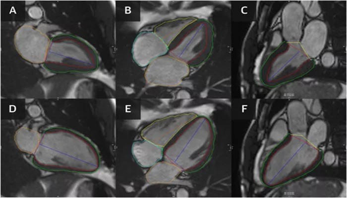
Magnetic Resonance Imaging (MRI) is a powerful, non-invasive diagnostic tool that plays a crucial role in modern medicine. It provides detailed images of the organs, tissues, and structures inside the body without the need for surgery or exposure to ionizing radiation, unlike X-rays and CT scans. MRI is widely used in the medical field, particularly for assessing the cardiovascular system, the brain, and musculoskeletal conditions.
MRI works by using a strong magnetic field, radio waves, and a computer to generate images. These images allow healthcare providers to diagnose a variety of medical conditions, monitor disease progression, and plan for treatments. In cardiovascular medicine, MRI is becoming increasingly valuable, particularly in diagnosing heart disease and evaluating its severity.
In this article, we will explore MRI in depth, covering its technology, how it works, the types of MRI scans available, and its applications, particularly in cardiovascular care. We will also discuss the benefits, risks, and considerations associated with MRI, as well as the advancements in the field.
How MRI Works: The Basics of Magnetic Resonance Imaging
MRI is based on a principle called nuclear magnetic resonance (NMR), which involves the interaction of nuclear spins with a magnetic field. The process begins when the patient is positioned inside a large magnet, which generates a strong magnetic field. This magnetic field causes the hydrogen nuclei in the body’s water molecules to align with the field.
Next, a radiofrequency pulse is applied, which temporarily disturbs the alignment of these hydrogen nuclei. When the pulse is turned off, the hydrogen atoms return to their original position, emitting energy in the form of signals. These signals are detected by the MRI machine and processed by a computer to create detailed images of the body’s internal structures.
Because the human body is primarily composed of water, which contains hydrogen atoms, MRI is particularly useful for imaging soft tissues such as the brain, heart, muscles, and organs. The resulting images are high in contrast, allowing for the visualization of minute details that might be missed by other imaging techniques.
Types of MRI Scans
MRI scans can be customized to target specific parts of the body or provide specific types of information. There are several variations of MRI, depending on the condition being assessed and the type of contrast agents used. The most common types of MRI include:
1. Structural MRI (Anatomical MRI)
Structural MRI is used to obtain detailed images of the body’s organs and tissues. It is often used in the evaluation of brain tumors, spinal cord abnormalities, joint and soft tissue injuries, and diseases like multiple sclerosis. This type of MRI provides high-resolution images of the body’s internal structures.
2. Functional MRI (fMRI)
Functional MRI, also known as fMRI, is used to measure and map brain activity. Unlike standard MRI, which focuses on the structure, fMRI detects changes in blood flow in the brain, which are linked to neural activity. It is commonly used in neuroscience research and in pre-surgical planning for brain surgery to map important brain functions like movement and speech.
3. Cardiovascular MRI (CMR)
Cardiovascular MRI, or CMR, is an advanced application of MRI that focuses on imaging the heart and blood vessels. CMR provides detailed images of the heart’s chambers, valves, and surrounding vessels, allowing doctors to assess heart function, detect heart disease, and evaluate the severity of conditions such as heart failure, congenital heart disease, coronary artery disease, and cardiomyopathy.
CMR also allows for tissue characterization, which is crucial in diagnosing myocardial infarction (heart attacks) and evaluating myocardial fibrosis (scarring in the heart muscle). The use of gadolinium-based contrast agents can enhance the imaging and provide further insights into tissue abnormalities.
4. Magnetic Resonance Angiography (MRA)
Magnetic Resonance Angiography is a specific MRI technique designed to visualize blood vessels. It is commonly used to diagnose conditions like aneurysms, arterial blockages, or abnormal blood flow in the brain, heart, or other organs. MRA can provide images of blood vessels without the need for invasive catheter procedures, making it a valuable tool in vascular diagnostics.
5. Diffusion Tensor Imaging (DTI)
Diffusion Tensor Imaging (DTI) is a specialized form of MRI that provides information about the orientation of white matter fibers in the brain. It is particularly useful in studying brain connectivity and detecting abnormalities in brain structures, such as those caused by stroke, traumatic brain injury, or neurological disorders.
Applications of MRI in Medicine
MRI has numerous applications in various medical specialties. Below are some of the most common uses for MRI:
1. Neurological Imaging
MRI is one of the primary imaging modalities used for diagnosing neurological conditions. It provides high-resolution images of the brain and spinal cord, making it essential for diagnosing brain tumors, stroke, multiple sclerosis, and degenerative conditions like Alzheimer’s disease and Parkinson’s disease. MRI also plays a crucial role in monitoring disease progression and planning treatments.
2. Musculoskeletal Imaging
MRI is invaluable for musculoskeletal imaging, particularly for soft tissues like muscles, tendons, and ligaments. It is commonly used to assess joint injuries, tears in the cartilage or ligaments, and conditions such as osteoarthritis. MRI is preferred over X-rays for soft tissue imaging, as it provides better contrast and clearer pictures.
3. Cardiovascular Imaging
Cardiovascular MRI (CMR) has become an important tool for diagnosing and assessing heart conditions. CMR can evaluate heart function, the size of the heart’s chambers, the blood flow through the heart, and the condition of the heart valves and blood vessels. It is used for diagnosing conditions such as coronary artery disease, heart failure, congenital heart defects, and valvular diseases.
4. Cancer Imaging
MRI is frequently used in oncology for detecting and staging various cancers. It is particularly useful in imaging soft tissue cancers, such as those in the brain, liver, breast, and prostate. MRI is also used to monitor the effectiveness of cancer treatments and detect any recurrence of tumors.
5. Abdominal Imaging
MRI can be used to assess a variety of abdominal conditions, including liver disease, kidney abnormalities, pancreatic disorders, and gastrointestinal conditions. Its ability to provide detailed images of soft tissues makes it a valuable tool for detecting tumors, cysts, and other abnormalities in the abdominal organs.
Benefits of MRI
MRI has several benefits that make it a preferred imaging tool in modern medicine:
Non-invasive: Unlike many other diagnostic tools, MRI does not require surgery or insertion of instruments into the body.
No radiation: MRI uses magnetic fields and radio waves, so there is no exposure to ionizing radiation, unlike X-rays or CT scans.
Detailed images: MRI provides high-resolution images of soft tissues, which allows healthcare providers to diagnose and evaluate conditions in greater detail.
Comprehensive diagnostic tool: MRI can be used to assess multiple areas of the body simultaneously, making it a valuable tool in the diagnosis and monitoring of various diseases.
Risks And Considerations
While MRI is generally safe, there are certain risks and considerations that need to be addressed:
Claustrophobia: MRI machines are typically enclosed, and some patients may experience anxiety or discomfort in the confined space. There are open MRI machines available for individuals who cannot tolerate the traditional closed MRI.
Metal implants: MRI uses powerful magnetic fields, so patients with certain metal implants (such as pacemakers, metal prostheses, or cochlear implants) may not be able to undergo MRI. Special precautions are taken in these cases, and some implants are MRI-compatible.
Contrast agents: Gadolinium-based contrast agents are commonly used in MRI to enhance image quality. While these agents are generally safe, they may cause allergic reactions in some individuals. People with kidney disease may need special consideration, as the contrast agent can cause complications in those with impaired kidney function.
Conclusion
Magnetic Resonance Imaging (MRI) is a powerful and versatile diagnostic tool that has transformed the way healthcare providers evaluate and treat patients. With its ability to provide high-resolution, detailed images of soft tissues and organs, MRI is invaluable in diagnosing a wide range of medical conditions, particularly in cardiology, neurology, musculoskeletal health, and cancer detection.
While MRI is generally safe and non-invasive, there are some risks and considerations that patients should be aware of, particularly with regard to metal implants and the use of contrast agents. However, with proper preparation and precautions, MRI remains one of the most reliable and effective imaging techniques in modern medicine, offering invaluable insights into the body’s internal structures and helping guide treatment decisions.
Related topics:


