Arrhythmias are disruptions in the normal rhythm of the heart, characterized by irregular heartbeats that can be too fast, too slow, or erratic. These irregularities can be fleeting or persistent, and while some are harmless, others can be life-threatening. Detecting arrhythmias is crucial for timely intervention, and one of the most effective tools for this purpose is the electrocardiogram (ECG or EKG). This article explores what arrhythmias are, how they manifest on an ECG, and the implications of these findings.
Understanding Arrhythmia
Arrhythmia refers to any disturbance in the heart’s rhythm. The heart’s electrical system controls the rate and rhythm of heartbeats. When this system malfunctions, it can lead to arrhythmias. There are several types of arrhythmias, including:
Bradycardia: A slower than normal heart rate.
Tachycardia: A faster than normal heart rate.
Atrial Fibrillation (AFib): Rapid, irregular beating of the atria.
Ventricular Fibrillation (VFib): Rapid, erratic beating of the ventricles.
Premature Beats: Extra beats that occur earlier than the next expected regular beat.
The Role of Electrocardiogram (ECG)
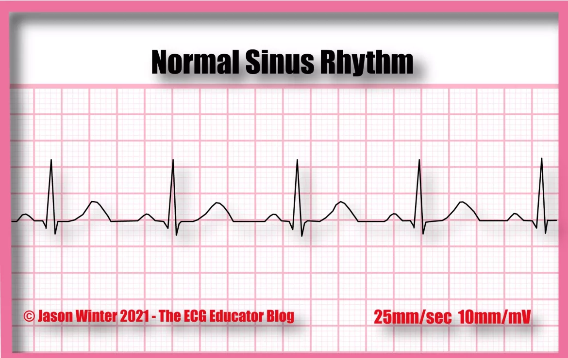
An ECG is a non-invasive test that records the electrical activity of the heart. It provides a visual representation of the heart’s electrical impulses as they travel through the heart muscle. These impulses are recorded as waves on a graph, each wave corresponding to a specific part of the heartbeat.
Key Components of An ECG
P Wave: Represents atrial depolarization, the first part of the heartbeat where the atria contract.
QRS Complex: Represents ventricular depolarization, where the ventricles contract.
T Wave: Represents ventricular repolarization, where the ventricles prepare for the next beat.
How Arrhythmias Appear on An ECG
Different types of arrhythmias produce characteristic changes in the ECG waveform. Here are some common patterns:
Bradycardia: A prolonged interval between the QRS complexes, indicating a slower heart rate.
Tachycardia: Shortened intervals between QRS complexes, indicating a faster heart rate.
Atrial Fibrillation: Absence of distinct P waves and an irregularly irregular rhythm.
Ventricular Fibrillation: Chaotic, irregular waveforms without discernible QRS complexes.
Premature Beats: Early occurrence of QRS complexes or P waves.
Types of Arrhythmias Detected by ECG
Sinus Arrhythmia
Sinus arrhythmia is a common and usually benign variation in heart rate that occurs with breathing. On an ECG, sinus arrhythmia is indicated by a variation in the R-R interval, which is the time between successive QRS complexes. This variation corresponds to the phases of respiration: heart rate typically increases during inhalation and decreases during exhalation.
Atrial Fibrillation (AFib)
Atrial fibrillation is characterized by rapid and irregular atrial contractions. On an ECG, AFib presents as an absence of discrete P waves before each QRS complex. Instead, there are erratic baseline waves and an irregularly irregular ventricular response. AFib is associated with an increased risk of stroke and requires medical attention for management and treatment.
Atrial Flutter
Atrial flutter is similar to AFib but has a more organized pattern. It appears on an ECG as a “sawtooth” pattern of flutter waves between QRS complexes. These flutter waves, or F waves, are more regular than the erratic waves seen in AFib. Atrial flutter can also lead to serious complications if not treated.
Ventricular Tachycardia (VT)
Ventricular tachycardia is a potentially life-threatening arrhythmia originating from the ventricles. On an ECG, VT is indicated by wide, bizarre-looking QRS complexes at a rapid rate, typically over 100 beats per minute. This condition can quickly deteriorate into ventricular fibrillation, which requires immediate medical intervention.
Ventricular Fibrillation (VFib)
Ventricular fibrillation is an emergency condition where the ventricles quiver ineffectively instead of contracting. The ECG shows a chaotic and irregular pattern without identifiable QRS complexes. VFib leads to a cessation of effective blood circulation and requires immediate defibrillation to prevent sudden cardiac death.
Premature Atrial and Ventricular Contractions (PACs and PVCs)
Premature contractions are early heartbeats that originate in the atria (PACs) or ventricles (PVCs). On an ECG, PACs appear as an early P wave followed by a normal QRS complex. PVCs appear as wide and bizarre QRS complexes, not preceded by a P wave. While often benign, frequent PACs or PVCs may require further investigation.
Diagnosis And Interpretation of Arrhythmias on ECG
The Importance of ECG in Arrhythmia Detection
An ECG is a primary tool for diagnosing arrhythmias due to its ability to provide real-time information about the heart’s electrical activity.
The test is quick, painless, and non-invasive, making it accessible for routine use. By analyzing the waveforms, healthcare providers can identify the type of arrhythmia, its severity, and potential underlying causes.
Clinical Implications of Arrhythmias Detected by ECG
Risk Stratification and Management
Identifying arrhythmias on an ECG is crucial for risk stratification and management. Certain arrhythmias, such as AFib, carry a high risk of stroke and require anticoagulation therapy. Others, like ventricular tachycardia, may necessitate implantable cardioverter-defibrillators (ICDs) to prevent sudden cardiac death.
Treatment Options
The treatment of arrhythmias detected by ECG varies depending on the type and severity. Options include:
Medications: Antiarrhythmic drugs, beta-blockers, and calcium channel blockers.
Electrical Cardioversion: A procedure to reset the heart’s rhythm using controlled electric shocks.
Catheter Ablation: A minimally invasive procedure to destroy the areas of the heart causing abnormal rhythms.
Pacemakers: Devices implanted to regulate slow heartbeats.
ICDs: Devices implanted to correct life-threatening arrhythmias by delivering shocks.
Conclusion
Arrhythmias are a group of conditions characterized by abnormal heart rhythms that can range from benign to life-threatening. The electrocardiogram (ECG) is an indispensable tool in the detection and diagnosis of these arrhythmias. By analyzing the electrical activity of the heart, healthcare providers can identify the type and severity of arrhythmias, guiding appropriate treatment and management strategies. Understanding arrhythmias and their detection through ECG is vital for preventing complications and ensuring optimal heart health.

