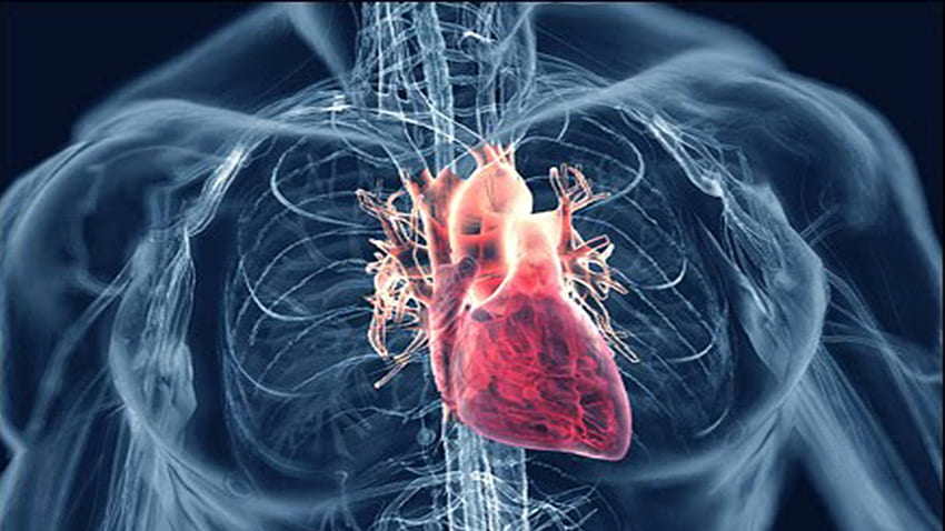
Heart failure is a complex clinical syndrome characterized by the inability of the heart to pump blood efficiently to meet the body’s demands. This condition can lead to a cascade of symptoms and signs, one of the most common being peripheral edema. Edema in heart failure occurs due to fluid retention, often accumulating in dependent areas of the body such as the legs, ankles, and abdomen. Understanding where to check for edema and how to interpret its presence is crucial for diagnosing and managing heart failure effectively.
Anatomy And Physiology of Edema in Heart Failure
Edema, simply put, is the abnormal accumulation of fluid in the interstitial spaces of tissues. In heart failure, several mechanisms contribute to the development of edema:
Increased Capillary Hydrostatic Pressure: As the heart’s pumping ability decreases, blood flow out of the heart is impaired, leading to increased pressure within the blood vessels (capillaries). This elevated pressure forces fluid out of the capillaries into the surrounding tissues, causing edema.
Fluid Retention and Sodium Retention: Heart failure often results in activation of the renin-angiotensin-aldosterone system (RAAS) and sympathetic nervous system, which leads to sodium and water retention by the kidneys. This fluid retention exacerbates the volume overload and contributes to edema formation.
Reduced Plasma Oncotic Pressure: In heart failure, there may be a decrease in plasma proteins (particularly albumin) due to liver dysfunction or malnutrition. Lower plasma oncotic pressure reduces the reabsorption of fluid from the tissues back into the capillaries, promoting edema.
SEE ALSO: How Heart Failure Causes Pleural Effusion
Where Should You Check for Edema Caused by Heart Failure
When assessing a patient for signs of heart failure, detecting and evaluating edema is a critical component. The clinical examination focuses on identifying the presence, extent, and characteristics of edema in various parts of the body. Here’s a systematic approach to evaluating edema caused by heart failure:
1. Lower Extremities
Peripheral edema is often the most noticeable and common manifestation of heart failure. It typically begins in the feet and ankles and may progress up the legs if the heart failure worsens. When assessing lower extremity edema, consider the following:
Location: Check for edema in the feet, ankles, shins, and thighs. Note any asymmetry between the legs.
Pitting Edema: Press firmly with your thumb over the bony prominences of the ankle or shin for 5 seconds and observe for the presence of pitting, which indicates the severity of fluid accumulation.
Skin Changes: Look for skin discoloration, shiny appearance, or warmth, which can indicate chronicity or inflammation associated with edema.
2. Sacral Area
In bedridden or elderly patients with heart failure, fluid may accumulate in the sacral area due to prolonged supine position and impaired circulation. Assess for sacral edema by:
Inspecting the Sacrum: Lift the patient’s gown or clothing to visually inspect the sacral area for swelling or fluid accumulation.
Palpation: Gently press on the sacral area to assess for pitting edema and note any skin changes.
3. Abdomen
Abdominal distension and fluid accumulation (ascites) can occur in patients with severe heart failure, particularly in cases involving right-sided heart failure or concomitant liver disease. Assess for abdominal edema by:
Inspection: Look for distension or bulging of the abdomen.
Palpation: Lightly palpate the abdomen to detect fluid waves or shifting dullness, which indicate the presence of ascites.
4. Lungs and Thorax
Pulmonary congestion is another hallmark of heart failure, where fluid accumulates in the lungs (pulmonary edema). While not directly visible externally, signs such as crackles on auscultation, shortness of breath, and orthopnea (breathing difficulties when lying flat) suggest fluid overload in the lungs.
Differential Diagnosis of Edema
When encountering edema, it’s essential to consider various differential diagnoses apart from heart failure. Conditions such as chronic venous insufficiency, nephrotic syndrome, liver cirrhosis, and medications (e.g., calcium channel blockers, NSAIDs) can also cause edema. Distinguishing between these etiologies is crucial for appropriate management.
Diagnostic Evaluation
Confirming the underlying cause of edema in suspected heart failure involves a comprehensive diagnostic approach:
Medical History and Physical Examination: Detailed history-taking and thorough physical examination focusing on cardiovascular and renal systems are essential.
Laboratory Tests: Blood tests including electrolytes, renal function tests, liver function tests, and brain natriuretic peptide (BNP) levels can provide valuable diagnostic information.
Imaging Studies: Chest X-ray and echocardiography are commonly used to assess cardiac structure and function, detect pulmonary congestion, and evaluate for signs of heart failure.
Management of Edema in Heart Failure
The management of edema in heart failure aims to alleviate symptoms, improve quality of life, and reduce hospitalizations.
Key principles include:
Diuretic Therapy: Loop diuretics (e.g., furosemide) are first-line agents to reduce fluid retention and relieve edema.
Sodium Restriction: Limiting dietary sodium intake helps to decrease fluid retention.
Fluid Restriction: In severe cases, restricting total fluid intake may be necessary to maintain fluid balance.
Compression Therapy: Use of compression stockings can help reduce lower extremity edema and improve venous return.
Elevating Legs: Advising patients to elevate their legs when sitting or lying down can assist in reducing dependent edema.
Conclusion
In conclusion, recognizing and assessing edema caused by heart failure is essential for diagnosing and managing this chronic condition effectively. By understanding the pathophysiology, conducting a thorough clinical evaluation, and employing appropriate diagnostic tools, healthcare providers can provide timely interventions to improve patient outcomes. Early recognition of edema allows for prompt initiation of treatment, which is crucial in mitigating symptoms and preventing disease progression in patients with heart failure.
Through a systematic approach to evaluating edema in various body regions—such as the lower extremities, sacral area, abdomen, and lungs—clinicians can gather valuable information to guide treatment decisions and optimize patient care. By addressing fluid overload and managing underlying cardiac dysfunction, healthcare teams can significantly enhance the quality of life for individuals living with heart failure.


