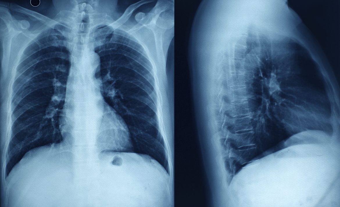
Myocarditis, an inflammatory condition of the heart muscle, poses diagnostic challenges due to its diverse clinical presentations and variable disease course. As a cardiologist specializing in arrhythmias, I frequently encounter cases where timely and accurate diagnosis of myocarditis is crucial for optimal management. This article aims to delve into the question: can chest X-rays show myocarditis? We will explore the diagnostic limitations and potential utility of chest X-rays in detecting myocarditis, alongside other imaging modalities and clinical considerations.
Understanding Myocarditis
Myocarditis refers to inflammation of the myocardium, typically caused by viral infections, autoimmune reactions, or exposure to toxins. The inflammatory process can lead to myocardial injury, dysfunction, and in severe cases, heart failure or arrhythmias. Clinical manifestations vary widely from mild flu-like symptoms to life-threatening cardiogenic shock or sudden cardiac death.
SEE ALSO: Does The J&J Vaccine Cause Myocarditis?
Clinical Presentation
The presentation of myocarditis can mimic other cardiac conditions or systemic illnesses, making its diagnosis challenging:
Symptoms: May include chest pain, shortness of breath, palpitations, fatigue, and signs of heart failure such as edema or fluid retention.
Clinical Signs: Physical examination findings such as abnormal heart sounds, murmurs, or evidence of fluid overload.
Diagnostic Challenges
Role of Imaging in Myocarditis
Imaging plays a critical role in the diagnostic workup of myocarditis, aiming to assess cardiac structure, function, and detect signs of inflammation or myocardial damage:
Echocardiography: Provides real-time imaging of the heart, assessing for chamber dimensions, wall motion abnormalities, and signs of myocardial dysfunction.
Cardiac Magnetic Resonance Imaging (MRI): Offers superior tissue characterization and is considered the gold standard for diagnosing myocarditis due to its ability to detect myocardial edema, inflammation, and scar tissue.
Nuclear Imaging (PET or SPECT): Can assess myocardial perfusion and inflammation, particularly useful in cases where MRI is contraindicated or inconclusive.
Limitations of Chest X-rays
While chest X-rays are routinely performed in patients with suspected cardiac pathology, their utility in diagnosing myocarditis is limited:
Imaging Features: Chest X-rays may show nonspecific findings such as cardiomegaly (enlarged heart), pulmonary congestion (evidence of fluid overload), or pleural effusions (fluid around the lungs), which can be secondary to heart failure but are not specific to myocarditis.
Sensitivity and Specificity: Chest X-rays lack sensitivity and specificity for detecting myocarditis directly. They may provide supportive evidence in the context of clinical suspicion and can help assess for complications such as pulmonary edema or effusions.
Pathophysiology of Myocarditis
Understanding the underlying pathophysiology of myocarditis helps elucidate why certain imaging modalities are more effective than others in its diagnosis:
Inflammatory Response: Myocarditis involves infiltration of immune cells into the myocardium, leading to cellular injury, edema, and potentially fibrosis.
Imaging Targets: MRI is sensitive to myocardial edema and inflammation, whereas chest X-rays primarily assess cardiac size and pulmonary status.
Clinical Approach to Diagnosis
Diagnostic Criteria
The diagnosis of myocarditis relies on a combination of clinical findings, biomarkers, imaging studies, and sometimes endomyocardial biopsy in select cases:
Biomarkers: Elevated levels of cardiac enzymes (e.g., troponin) and inflammatory markers (e.g., C-reactive protein) may support the diagnosis.
Imaging Modalities: MRI findings of myocardial edema (T2-weighted imaging) and late gadolinium enhancement (indicative of fibrosis or scar) are characteristic but require expertise in interpretation.
Role of Chest X-rays
In clinical practice, chest X-rays are typically part of the initial evaluation in patients presenting with cardiac symptoms or signs of heart failure:
Detection of Complications: Chest X-rays can identify complications such as pulmonary congestion or pleural effusions, which may occur secondary to myocarditis-induced heart failure.
Supportive Evidence: While chest X-rays do not directly visualize myocardial inflammation, they can provide indirect evidence of cardiac involvement and guide further diagnostic steps.
Clinical Scenarios
Case Studies
Consider the following scenarios where chest X-rays may play a role in the diagnostic pathway of myocarditis:
Case 1: A young adult presents with chest pain, fever, and elevated cardiac biomarkers. Chest X-ray reveals cardiomegaly and pulmonary congestion, prompting further evaluation with echocardiography and cardiac MRI.
Case 2: An elderly patient with known coronary artery disease presents with worsening heart failure symptoms. Chest X-ray shows bilateral pleural effusions and signs of pulmonary edema. Clinical suspicion for myocarditis arises, leading to comprehensive imaging and laboratory workup.
Conclusion
In conclusion, while chest X-rays are valuable in assessing cardiac size and detecting secondary pulmonary changes, their utility in directly diagnosing myocarditis is limited. The diagnosis of myocarditis requires a comprehensive approach integrating clinical assessment, biomarkers, and advanced imaging modalities such as cardiac MRI for accurate evaluation of myocardial inflammation and injury.


