The heart, a vital organ responsible for pumping blood throughout the body, is nourished by a network of coronary arteries.
These arteries supply oxygen-rich blood to the heart muscle, ensuring its proper function. Among these arteries, the right coronary artery (RCA) plays a crucial role. Understanding the location and function of the RCA is essential for comprehending heart anatomy and the mechanisms of various cardiovascular diseases.
Anatomy of The Coronary Arteries
The coronary arteries are classified into two main branches: the left coronary artery (LCA) and the right coronary artery (RCA). Each of these arteries further branches out to supply different regions of the heart.
Left Coronary Artery (LCA): This artery divides into the left anterior descending (LAD) artery and the left circumflex (LCx) artery. The LAD supplies the front and bottom of the left side of the heart, while the LCx supplies the left atrium and the side and back of the left ventricle.
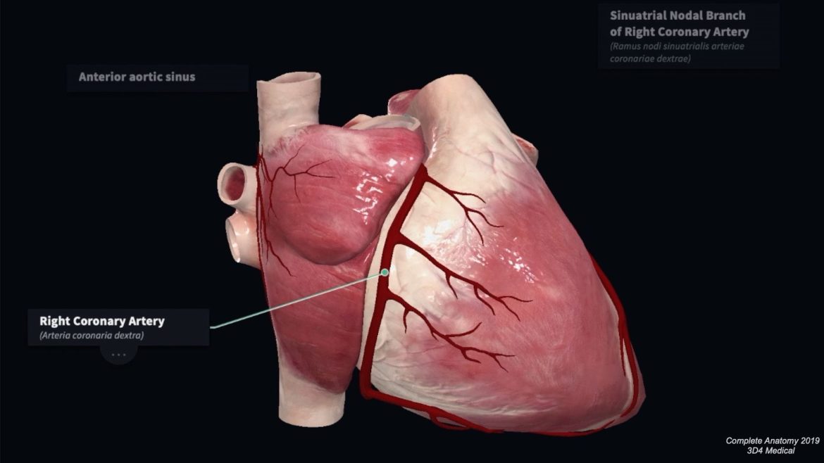
Right Coronary Artery (RCA): This artery supplies the right atrium, right ventricle, bottom portion of both ventricles, and the back of the septum.
SEE ALSO: How to Diagnose Acute Coronary Syndrome
Where Is The Right Coronary Artery Located in The Heart?
The right coronary artery originates from the right aortic sinus, a region in the aorta just above the aortic valve. It travels along the right atrioventricular (AV) groove, a groove between the right atrium and right ventricle. The RCA then descends toward the back of the heart, giving off several important branches.
Branches of the Right Coronary Artery
The RCA has several branches that supply various parts of the heart.
These include:
Right Marginal Artery: This artery branches off the RCA and travels along the lower border of the heart, supplying the right ventricle.
Posterior Descending Artery (PDA): Also known as the posterior interventricular artery, the PDA runs along the posterior interventricular sulcus and supplies the posterior third of the interventricular septum.
Atrial Branches: These small branches supply the right atrium.
Conus Artery: This artery supplies the conus arteriosus (infundibulum), the outflow tract of the right ventricle.
Function of The Right Coronary Artery
Oxygen Supply to the Heart Muscle
The primary function of the RCA is to deliver oxygen-rich blood to the heart muscle (myocardium). By supplying the right atrium, right ventricle, bottom part of the left ventricle, and the back of the septum, the RCA ensures that these areas receive the necessary oxygen and nutrients to function effectively.
Electrical Conduction System
The RCA also plays a crucial role in the heart’s electrical conduction system. It supplies blood to the sinoatrial (SA) node in approximately 60% of individuals and the atrioventricular (AV) node in about 85-90% of individuals. These nodes are essential for maintaining the heart’s rhythm.
Clinical Significance
Coronary Artery Disease
Coronary artery disease (CAD) is a condition characterized by the narrowing or blockage of the coronary arteries due to plaque buildup.
When the RCA is affected by CAD, it can lead to a reduction in blood flow to the areas it supplies, potentially causing ischemia and myocardial infarction (heart attack).
Symptoms of RCA Blockage
Blockage of the RCA can result in symptoms such as:
- Chest pain (angina)
- Shortness of breath
- Fatigue
- Dizziness
- Heart palpitations
Diagnosis of RCA Disease
Several diagnostic tests can help identify issues with the RCA, including:
Electrocardiogram (ECG): This test measures the electrical activity of the heart and can indicate areas of ischemia or infarction.
Stress Test: This test evaluates the heart’s function under stress, often revealing reduced blood flow in the RCA.
Coronary Angiography: This imaging technique uses contrast dye and X-rays to visualize the coronary arteries and identify blockages.
CT Coronary Angiography: A non-invasive imaging test that provides detailed images of the coronary arteries.
Treatment Options
Treatment for RCA disease may include lifestyle changes, medications, and surgical interventions. These can include:
Lifestyle Changes: Diet, exercise, and smoking cessation can help improve heart health.
Medications: Drugs such as statins, beta-blockers, and antiplatelet agents can manage symptoms and reduce the risk of complications.
Percutaneous Coronary Intervention (PCI): Also known as angioplasty, this procedure involves inflating a balloon inside the artery to widen it and placing a stent to keep it open.
Coronary Artery Bypass Grafting (CABG): This surgical procedure creates a new pathway for blood to flow around the blocked artery.
Variations in RCA Anatomy
Anatomical Variations
The RCA can exhibit variations in its anatomy, which can affect its branching patterns and the areas it supplies. These variations can be significant in clinical settings, particularly during surgical procedures and diagnostic evaluations.
Dominance Patterns
The coronary artery system can be classified based on the dominance pattern, which refers to which artery supplies the posterior descending artery (PDA). The three main patterns are:
Right-Dominant: The RCA supplies the PDA. This is the most common pattern, occurring in about 70-85% of individuals.
Left-Dominant: The left circumflex artery (LCx) supplies the PDA. This occurs in about 8-10% of individuals.
Co-Dominant: Both the RCA and the LCx contribute to the supply of the PDA. This pattern is seen in about 7-20% of individuals.
Imaging And Diagnostic Techniques
Invasive Imaging
Coronary Angiography is the gold standard for visualizing the coronary arteries. This procedure involves the insertion of a catheter into the coronary arteries and the injection of contrast dye to highlight the arteries on X-ray images.
Non-Invasive Imaging
Non-invasive imaging techniques are also used to assess the RCA and its branches. These include:
Computed Tomography Coronary Angiography (CTCA): This imaging test uses CT scans to create detailed images of the coronary arteries.
Magnetic Resonance Angiography (MRA): This technique uses magnetic fields and radio waves to visualize the coronary arteries.
Functional Testing
Functional tests assess the impact of RCA disease on the heart’s function. These tests include:
Stress Echocardiography: This test combines an exercise stress test with an echocardiogram to evaluate heart function under stress.
Nuclear Stress Test: This test uses a small amount of radioactive material to visualize blood flow to the heart during stress.
Advances in RCA Research
Innovations in Imaging
Advancements in imaging technology have significantly improved the visualization and assessment of the RCA. Techniques such as 3D coronary angiography and advanced CT imaging provide more detailed and accurate images, aiding in diagnosis and treatment planning.
Genetic Research
Genetic research has identified specific genetic markers associated with an increased risk of coronary artery disease, including RCA disease. Understanding these genetic factors can lead to more personalized treatment and prevention strategies.
Interventional Techniques
Innovations in interventional techniques, such as drug-eluting stents and bioresorbable vascular scaffolds, have improved the outcomes of PCI procedures. These advancements reduce the risk of restenosis (re-narrowing of the artery) and improve long-term results.
Conclusion
The right coronary artery is a vital component of the heart’s circulatory system, supplying oxygen-rich blood to essential areas of the heart. Its location, branching patterns, and function are crucial for maintaining heart health. Understanding the RCA’s anatomy and its role in cardiovascular disease is essential for diagnosing and treating heart conditions effectively.
Ongoing research and technological advancements continue to enhance our knowledge and capabilities in managing RCA-related diseases, ultimately improving patient outcomes and quality of life.

