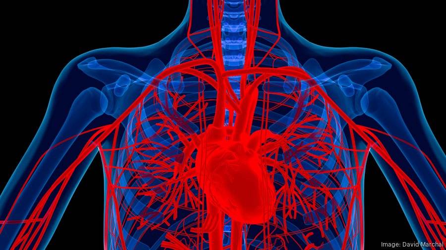
Ventricular triad refers to a specific set of three clinical findings typically observed in the context of ventricular arrhythmias, especially in severe cardiac conditions. These findings usually include hypotension (low blood pressure), distended neck veins, and muffled heart sounds. While ventricular triad is often associated with cardiac tamponade—a life-threatening condition where fluid accumulates in the pericardium, exerting pressure on the heart—this article will explore the broader causes of ventricular triad, focusing on its origins, risk factors, and underlying pathophysiology.
What Are The Causes of Ventricular Triad?
Cardiac Tamponade
Pathophysiology and Clinical Presentation
Cardiac tamponade is one of the primary conditions associated with ventricular triad. This condition occurs when fluid, blood, or gas accumulates in the pericardial space, creating pressure on the heart and restricting its normal filling and function. The resultant compression leads to a decrease in cardiac output and subsequently, hypotension. The distended neck veins result from elevated venous pressure as blood return to the heart is impaired. Muffled heart sounds are due to the insulating effect of the accumulated fluid around the heart.
SEE ALSO: 6 Treatments for Systolic Hypertension
Causes of Cardiac Tamponade
Trauma: Blunt or penetrating chest trauma can lead to bleeding into the pericardial space, causing tamponade.
Pericarditis: Inflammation of the pericardium, often due to infections (viral, bacterial, or fungal), autoimmune conditions, or post-myocardial infarction syndrome, can result in fluid accumulation.
Malignancies: Metastatic cancers, especially lung, breast, and hematological cancers, can invade the pericardium, leading to fluid build-up.
Uremia: Chronic kidney disease can lead to pericarditis and subsequent tamponade due to metabolic waste accumulation.
Medical Procedures: Complications from cardiac surgeries, catheter insertions, or pacemaker placements can result in accidental bleeding into the pericardial space.
Myocardial Infarction
Pathophysiology and Clinical Presentation
Acute myocardial infarction (MI), commonly known as a heart attack, can also present with signs resembling the ventricular triad. The damage to the myocardial tissue can impair the heart’s pumping ability, leading to hypotension. In severe cases, particularly with right ventricular infarction, distended neck veins can be observed due to increased venous pressure. Additionally, the heart sounds might be muffled due to complications like pericardial effusion or ventricular septal rupture.
Causes of Myocardial Infarction
Atherosclerosis: The primary cause of MI is the occlusion of coronary arteries due to atherosclerotic plaque rupture and subsequent thrombus formation.
Coronary Artery Spasm: Spasms of the coronary arteries, often related to substance abuse (e.g., cocaine), can transiently block blood flow, leading to MI.
Emboli: Embolization of thrombi, vegetations from endocarditis, or other material into the coronary arteries can cause infarction.
Coronary Artery Dissection: A tear in the coronary artery wall can impede blood flow and result in MI.
Pulmonary Embolism
Pathophysiology and Clinical Presentation
Pulmonary embolism (PE) can sometimes mimic the signs of ventricular triad, particularly in massive PE. The obstruction of the pulmonary arteries increases the pressure in the right ventricle, leading to distended neck veins. The resultant strain on the heart can reduce cardiac output, causing hypotension. Muffled heart sounds may be present due to associated pericardial effusion or reduced cardiac function.
Causes of Pulmonary Embolism
Deep Vein Thrombosis (DVT): The most common cause, where thrombi from the deep veins of the legs travel to the lungs.
Hypercoagulable States: Conditions like cancer, pregnancy, and certain genetic disorders increase the risk of clot formation.
Immobility: Prolonged bed rest, long flights, or surgeries can lead to blood stasis and clot formation.
Trauma or Surgery: Injuries or surgical procedures, particularly orthopedic surgeries, can predispose individuals to PE.
Constrictive Pericarditis
Pathophysiology and Clinical Presentation
Constrictive pericarditis is a chronic condition where the pericardium becomes thickened and rigid, restricting the heart’s normal expansion and contraction. This restriction leads to symptoms similar to ventricular triad due to impaired cardiac filling and output. Distended neck veins and hypotension are common, and muffled heart sounds can occur due to the thickened pericardial sac.
Causes of Constrictive Pericarditis
Tuberculosis: Historically a common cause, particularly in regions where TB is prevalent.
Radiation Therapy: Treatment for cancers, especially those involving the chest, can lead to pericardial scarring.
Surgery: Previous cardiac surgeries can result in pericardial damage and subsequent constriction.
Connective Tissue Disorders: Diseases like rheumatoid arthritis and systemic lupus erythematosus can involve the pericardium.
Advanced Heart Failure
Pathophysiology and Clinical Presentation
Severe or advanced heart failure, particularly in the decompensated state, can present with signs similar to ventricular triad.
The inability of the heart to pump effectively leads to hypotension and elevated venous pressure, causing distended neck veins. Heart sounds might be muffled due to fluid overload and associated pericardial effusion.
Causes of Advanced Heart Failure
Ischemic Heart Disease: Chronic damage from coronary artery disease and repeated myocardial infarctions.
Cardiomyopathies: Dilated, hypertrophic, and restrictive cardiomyopathies can all progress to heart failure.
Hypertension: Long-standing high blood pressure leads to left ventricular hypertrophy and eventual heart failure.
Valvular Heart Disease: Conditions like aortic stenosis, mitral regurgitation, and others can culminate in heart failure.
Aortic Dissection
Pathophysiology and Clinical Presentation
Aortic dissection, particularly involving the ascending aorta, can present with features of the ventricular triad if it leads to pericardial tamponade. The tear in the aortic wall allows blood to enter the pericardial space, causing pressure on the heart.
Hypotension, distended neck veins, and muffled heart sounds are indicative of this catastrophic event.
Causes of Aortic Dissection
Hypertension: The most significant risk factor, with chronic high blood pressure contributing to the weakening of the aortic wall.
Connective Tissue Disorders: Conditions like Marfan syndrome and Ehlers-Danlos syndrome predispose individuals to dissection.
Atherosclerosis: Plaque buildup can weaken the aortic wall, making it susceptible to dissection.
Trauma: Severe chest trauma, including from car accidents, can cause aortic dissection.
Pericardial Effusion
Pathophysiology and Clinical Presentation
Pericardial effusion, the accumulation of fluid within the pericardial cavity, can result in tamponade and present with ventricular triad. The fluid buildup restricts the heart’s ability to expand fully, leading to hypotension, distended neck veins, and muffled heart sounds.
Causes of Pericardial Effusion
Infections: Viral, bacterial, and fungal infections can lead to pericardial effusion.
Autoimmune Diseases: Conditions like lupus and rheumatoid arthritis can cause inflammation and fluid accumulation.
Malignancies: Cancerous involvement of the pericardium can result in effusion.
Post-Cardiac Injury: Following myocardial infarction or cardiac surgery, inflammation and effusion can occur.
Conclusion
The ventricular triad, characterized by hypotension, distended neck veins, and muffled heart sounds, is a clinical finding indicative of significant underlying cardiac pathology. While commonly associated with cardiac tamponade, this triad can result from various conditions, including myocardial infarction, pulmonary embolism, constrictive pericarditis, advanced heart failure, aortic dissection, and pericardial effusion. Understanding the diverse causes and pathophysiological mechanisms behind ventricular triad is crucial for prompt diagnosis and effective management, ultimately improving patient outcomes in these potentially life-threatening situations.

