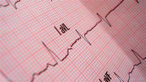
Arrhythmias, or irregular heartbeats, are conditions that can vary widely in their presentation and severity. They can range from harmless, momentary disruptions to life-threatening anomalies requiring immediate medical intervention. For medical professionals and individuals with heart conditions, understanding what arrhythmia looks like on a heart monitor is crucial. This article delves into the types of arrhythmias, how they appear on electrocardiograms (ECGs or EKGs), and the implications of these readings.
The Basics of Heart Monitors
Heart monitors, particularly electrocardiograms, are essential tools in cardiology. They record the electrical activity of the heart over a period of time and produce a series of waveforms that represent the heart’s rhythm and electrical conduction. A standard ECG tracing has several key components:
P wave: Represents atrial depolarization, the initial phase of the heartbeat where the atria contract.
QRS complex: Represents ventricular depolarization, the main pumping action where the ventricles contract.
T wave: Represents ventricular repolarization, where the ventricles prepare for the next contraction.
PR interval: The time between the start of atrial depolarization and the start of ventricular depolarization.
QT interval: The time it takes for the ventricles to depolarize and then repolarize.
SEE ALSO: What Will Be The Prevalence of Cardiac Arrhythmias by 2024?
Normal Sinus Rhythm
In a healthy heart, the ECG shows a consistent and predictable pattern known as the normal sinus rhythm. This rhythm is characterized by regular P waves, followed by QRS complexes and T waves in a predictable sequence. The heart rate typically ranges from 60 to 100 beats per minute (bpm) in adults, with equal intervals between each beat.
Types of Arrhythmias And Their ECG Presentations
1. Atrial Fibrillation (AFib)
Atrial fibrillation is one of the most common types of arrhythmia, where the atria beat irregularly and out of coordination with the ventricles. On an ECG, AFib is identified by the absence of distinct P waves and the presence of irregularly spaced QRS complexes. Instead of the regular P wave, the baseline appears wavy and chaotic, reflecting the rapid and disorganized electrical activity in the atria.
2. Atrial Flutter
Atrial flutter is characterized by a rapid, but regular, atrial rhythm. The ECG shows a “sawtooth” pattern of P waves, known as flutter waves, between each QRS complex. The ventricular rate may be regular or irregular, depending on how many of these flutter waves are conducted through the AV node to the ventricles.
3. Supraventricular Tachycardia (SVT)
SVT is a broad term that encompasses several types of arrhythmias originating above the ventricles. On an ECG, SVT is identified by a rapid heart rate, often between 150 and 250 bpm, with narrow QRS complexes. P waves may be difficult to distinguish because they can be buried in the preceding T wave due to the rapid rate.
4. Ventricular Tachycardia (VT)
Ventricular tachycardia is a serious arrhythmia that originates in the ventricles. On an ECG, VT is characterized by a regular, rapid heart rate, typically between 100 and 250 bpm, with wide and abnormal QRS complexes. The P waves are often absent or not associated with the QRS complexes.
5. Ventricular Fibrillation (VFib)
Ventricular fibrillation is a life-threatening condition where the ventricles quiver ineffectively instead of contracting properly. The ECG shows a chaotic, irregular waveform without any discernible QRS complexes, P waves, or T waves.
Immediate medical intervention with defibrillation is required to restore a normal rhythm.
6. Bradyarrhythmias
Bradyarrhythmias are slow heart rhythms, typically less than 60 bpm.
They can arise from issues with the heart’s natural pacemaker (the sinoatrial node) or the conduction pathways. On an ECG, bradyarrhythmias are identified by prolonged intervals between QRS complexes. Specific types include:
Sinus Bradycardia: A slow, but otherwise normal, rhythm with regular P waves and QRS complexes.
Heart Block: There are different degrees of heart block, each with distinct ECG features. For example, a first-degree block shows a prolonged PR interval, while a third-degree block (complete heart block) shows a complete dissociation between P waves and QRS complexes.
Recognizing Arrhythmias in Real-Time Monitoring
Holter Monitors and Event Recorders
For patients with intermittent arrhythmias, a standard ECG might not capture the irregular activity. In such cases, Holter monitors and event recorders are used. Holter monitors continuously record the heart’s activity over 24 to 48 hours, while event recorders are used for longer periods and can be triggered by the patient during symptoms. These devices provide a comprehensive picture of the heart’s rhythm over time.
Implantable Loop Recorders
Implantable loop recorders (ILRs) are used for long-term monitoring, often up to three years. They are implanted under the skin and continuously monitor the heart’s electrical activity, automatically recording any abnormal rhythms. ILRs are particularly useful for diagnosing unexplained syncope (fainting) or infrequent arrhythmias.
Clinical Implications of Arrhythmia Detection
Diagnosis and Treatment Planning
Detecting and interpreting arrhythmias on a heart monitor is critical for accurate diagnosis and treatment planning. For instance, distinguishing between atrial fibrillation and atrial flutter can guide the choice of anticoagulant therapy and rhythm control strategies.
Similarly, identifying life-threatening arrhythmias like ventricular fibrillation can prompt immediate life-saving interventions.
Risk Stratification
ECG findings help in risk stratification of patients. For example, the presence of frequent premature ventricular contractions (PVCs) or non-sustained ventricular tachycardia in a patient with structural heart disease might indicate a higher risk of sudden cardiac arrest, warranting more aggressive monitoring and treatment.
Guiding Interventions
Real-time monitoring and analysis of arrhythmias can guide interventions such as catheter ablation, where abnormal electrical pathways in the heart are targeted and destroyed. Precise localization of the arrhythmia origin is essential for the success of these procedures.
Patient Education and Management
For patients, understanding their arrhythmia and recognizing symptoms is crucial for effective management. Educating patients on how to use portable ECG devices or mobile apps that record heart rhythms can empower them to take an active role in their healthcare.
Advances in Arrhythmia Detection
Wearable Technology
Recent advances in wearable technology have made continuous heart monitoring more accessible. Devices like smartwatches and fitness trackers now come with ECG capabilities, allowing users to monitor their heart rhythm in real-time and detect arrhythmias. These technologies can alert users to seek medical attention if an irregular rhythm is detected.
Artificial Intelligence and Machine Learning
Artificial intelligence (AI) and machine learning algorithms are being integrated into ECG interpretation to enhance accuracy and speed.
These technologies can analyze vast amounts of data to identify subtle patterns and predict the likelihood of arrhythmias, aiding clinicians in early diagnosis and intervention.
Conclusion
Understanding what arrhythmia looks like on a heart monitor is essential for both medical professionals and patients. ECGs provide a visual representation of the heart’s electrical activity, allowing for the identification of various arrhythmias and guiding appropriate treatment. With advances in technology, continuous monitoring and early detection of arrhythmias are becoming more feasible, ultimately improving patient outcomes. Recognizing the signs of arrhythmia on a heart monitor and understanding their clinical implications can lead to timely and effective management of these conditions.

