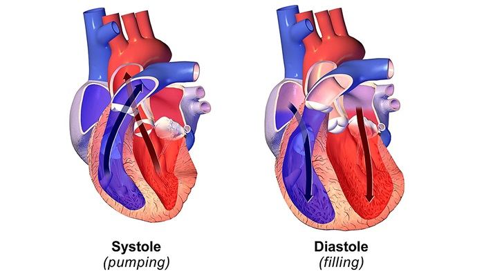
Diastolic dysfunction is a condition where the heart’s ability to relax and fill with blood during the diastolic phase is impaired. This can lead to heart failure with preserved ejection fraction (HFpEF), a condition that affects many individuals, particularly older adults. Accurate diagnosis of diastolic dysfunction is crucial for effective management and treatment. In this article, we will explore the 4 best ways to diagnose diastolic dysfunction, providing an in-depth look at each method, its procedures, advantages, and limitations.
1. Echocardiography
Echocardiography is a cornerstone in the diagnosis of diastolic dysfunction. This non-invasive imaging technique uses ultrasound waves to create detailed images of the heart, allowing clinicians to assess its structure and function in real-time.
Procedure
During an echocardiogram, a transducer is placed on the patient’s chest, emitting high-frequency sound waves that bounce off the heart’s structures. These echoes are captured and translated into images by the echocardiography machine. The procedure typically involves both M-mode and Doppler echocardiography:
M-mode Echocardiography: Provides a single-dimensional view of the heart, useful for measuring chamber size and wall thickness.
Doppler Echocardiography: Assesses blood flow through the heart’s chambers and valves, which is critical for evaluating diastolic function.
see also: What Is The Number One Drug for Heart Failure?
Key Measurements
Several echocardiographic parameters are used to diagnose diastolic dysfunction:
E/A Ratio: The ratio of early (E) to late (A) ventricular filling velocities obtained from pulsed-wave Doppler across the mitral valve. A reduced E/A ratio suggests impaired relaxation.
Deceleration Time (DT): The time it takes for the early diastolic flow to decelerate, with prolonged DT indicating diastolic dysfunction.
Tissue Doppler Imaging (TDI): Measures the velocity of myocardial motion, particularly the early diastolic (e’) and late diastolic (a’) velocities at the mitral annulus. A reduced e’ velocity is indicative of impaired relaxation.
Left Atrial Volume Index (LAVI): Increased left atrial size often reflects chronic diastolic dysfunction and elevated filling pressures.
Advantages
Non-invasive: Echocardiography is a non-invasive, painless procedure.
Comprehensive Assessment: Provides detailed information on both systolic and diastolic function, heart structure, and valvular abnormalities.
Widespread Availability: Echocardiography is widely available and can be performed in various healthcare settings.
Limitations
Operator Dependent: The accuracy of the results can be influenced by the skill and experience of the operator.
Variable Interpretations: Differences in measurement techniques and interpretations can lead to variability in diagnosing diastolic dysfunction.
2. Cardiac Magnetic Resonance Imaging (CMR)
Cardiac Magnetic Resonance Imaging (CMR) is another powerful tool for diagnosing diastolic dysfunction. CMR provides high-resolution images and precise quantification of cardiac function and tissue characteristics.
Procedure
CMR involves the use of a strong magnetic field and radio waves to produce detailed images of the heart. Patients lie within the MRI scanner, and various sequences are performed to evaluate different aspects of cardiac function and structure. Key CMR techniques relevant to diastolic dysfunction include:
Cine MRI: Produces moving images of the heart, allowing assessment of ventricular volume, mass, and wall motion.
T1 and T2 Mapping: Provides information on tissue composition, helping identify fibrosis or other myocardial abnormalities.
Phase Contrast Imaging: Measures blood flow velocities, similar to Doppler echocardiography, but with higher precision.
Key Measurements
Ventricular Volumes and Mass: Accurate quantification of ventricular volumes and mass helps in assessing overall cardiac function.
Myocardial Strain Analysis: Evaluates myocardial deformation, providing insights into subtle dysfunction that may not be apparent with conventional measurements.
Late Gadolinium Enhancement (LGE): Identifies areas of fibrosis or scarring within the myocardium, which can be associated with diastolic dysfunction.
Advantages
High Resolution: CMR provides superior image resolution and detailed tissue characterization.
Reproducibility: High reproducibility of measurements, making it a reliable tool for longitudinal studies.
Comprehensive Assessment: CMR can assess not only diastolic function but also detect underlying myocardial diseases.
Limitations
Cost and Accessibility: CMR is more expensive and less widely available compared to echocardiography.
Contraindications: Patients with certain implants or claustrophobia may not be suitable for CMR.
Time-Consuming: The procedure takes longer than an echocardiogram and may require the use of contrast agents.
3. Invasive Hemodynamic Monitoring
Invasive hemodynamic monitoring is considered the gold standard for the diagnosis of diastolic dysfunction, particularly in complex cases where non-invasive methods are inconclusive. This approach involves direct measurement of intracardiac pressures through cardiac catheterization.
Procedure
Cardiac catheterization involves inserting a catheter into a blood vessel, usually in the groin or arm, and guiding it to the heart. Once in place, pressure measurements are taken from different chambers of the heart. Key measurements for assessing diastolic function include:
Left Ventricular End-Diastolic Pressure (LVEDP): Elevated LVEDP is a hallmark of diastolic dysfunction.
Pulmonary Capillary Wedge Pressure (PCWP): Reflects left atrial pressure and can indicate elevated filling pressures.
Pressure-Volume Loops: Provide a detailed assessment of ventricular compliance and stiffness.
Advantages
Direct Measurement: Provides direct and precise measurement of intracardiac pressures, essential for accurate diagnosis.
Detailed Hemodynamic Assessment: Allows for a comprehensive understanding of cardiac function and hemodynamics.
Limitations
Invasive: The procedure carries risks associated with invasive techniques, including bleeding, infection, and vascular complications.
Cost and Resources: Requires specialized equipment and skilled personnel, making it more resource-intensive.
Patient Discomfort: Involves discomfort for the patient due to the invasive nature of the procedure.
4. Biomarkers
Biomarkers are increasingly being used as adjuncts in the diagnosis of diastolic dysfunction. These blood tests can provide insights into the underlying pathophysiology and severity of the condition.
Key Biomarkers
B-type Natriuretic Peptide (BNP) and N-terminal pro-BNP (NT-proBNP): Elevated levels of these peptides are indicative of increased cardiac stress and are commonly elevated in diastolic dysfunction.
Galectin-3: Associated with fibrosis and inflammation, elevated levels can indicate adverse cardiac remodeling.
ST2: A marker of myocardial stress and fibrosis, elevated ST2 levels are linked to worse outcomes in heart failure patients.
Advantages
Non-invasive: Biomarker testing is a simple blood test, making it non-invasive and easy to perform.
Early Detection: Can help in the early detection of diastolic dysfunction before significant symptoms develop.
Prognostic Value: Provides prognostic information, helping to stratify patients based on their risk of adverse outcomes.
Limitations
Non-specific: Elevated levels can be seen in various cardiac and non-cardiac conditions, reducing specificity for diastolic dysfunction.
Variability: Biomarker levels can be influenced by age, renal function, and other comorbidities, leading to variability in interpretation.
Adjunct Role: Biomarkers are best used in conjunction with imaging and clinical assessment rather than as standalone diagnostic tools.
Conclusion
Diagnosing diastolic dysfunction requires a multifaceted approach, combining clinical evaluation with advanced diagnostic techniques. Echocardiography, Cardiac Magnetic Resonance Imaging (CMR), Invasive Hemodynamic Monitoring, and Biomarkers each offer unique advantages and insights, making them the 4 best ways to diagnose diastolic dysfunction.
Understanding the strengths and limitations of each method allows clinicians to make informed decisions, ensuring accurate diagnosis and optimal management for patients with diastolic dysfunction.

