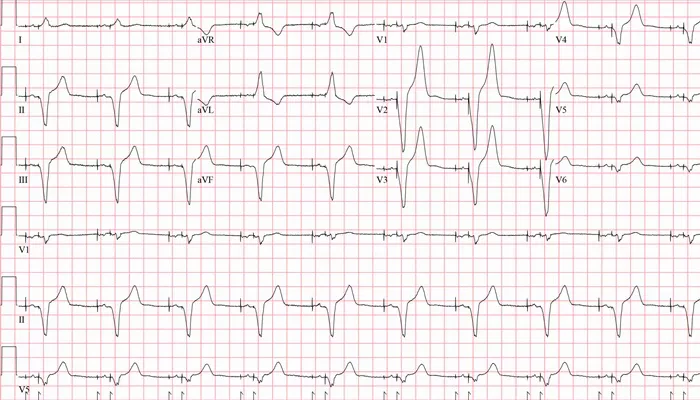
Understanding how to read arrhythmia strips is crucial for healthcare professionals, particularly cardiologists, as it enables them to accurately diagnose and treat various heart conditions. This article will guide you through the process of reading arrhythmia strips, covering the basics of electrocardiograms (ECGs), identifying different types of arrhythmias, and interpreting key ECG components.
Introduction to Electrocardiograms (ECGs)
An electrocardiogram (ECG) is a non-invasive diagnostic tool used to record the electrical activity of the heart. It provides valuable information about the heart’s rhythm and electrical conduction, helping to identify abnormalities such as arrhythmias. ECGs are typically displayed as a series of waves on graph paper, with each wave representing a specific aspect of the heart’s electrical cycle.
SEE ALSO: What Is SVT Arrhythmia?
Components of An ECG Strip
Understanding the components of an ECG strip is essential for accurate interpretation. The main components include:
P Wave: Represents atrial depolarization.
QRS Complex: Represents ventricular depolarization.
T Wave: Represents ventricular repolarization.
PR Interval: Measures the time from the onset of atrial depolarization to the onset of ventricular depolarization.
QT Interval: Measures the total time of ventricular depolarization and repolarization.
Steps to Read Arrhythmia Strips
Step 1: Assess the Rhythm
The first step in reading an arrhythmia strip is to assess the rhythm. Determine if the rhythm is regular or irregular by measuring the intervals between R waves (the peaks of the QRS complex). Regular intervals indicate a regular rhythm, while varying intervals suggest an irregular rhythm.
Step 2: Measure the Heart Rate
Next, measure the heart rate by counting the number of R waves in a 6-second strip and multiplying by 10. This gives the heart rate in beats per minute (bpm). Alternatively, you can use the 1500 method: count the number of small squares between two consecutive R waves and divide 1500 by this number.
Step 3: Evaluate the P Waves
Examine the P waves to ensure they are present and consistent before each QRS complex. Assess their shape and duration, which should be less than 0.12 seconds. P wave abnormalities can indicate atrial arrhythmias.
Step 4: Measure the PR Interval
Measure the PR interval from the beginning of the P wave to the start of the QRS complex. The normal PR interval ranges from 0.12 to 0.20 seconds. A prolonged PR interval suggests a first-degree heart block, while a short PR interval can indicate pre-excitation syndromes like Wolff-Parkinson-White syndrome.
Step 5: Assess the QRS Complex
Evaluate the QRS complex’s duration and morphology. A normal QRS duration is less than 0.12 seconds. A wide QRS complex may indicate a bundle branch block or ventricular arrhythmia. Additionally, assess for abnormal Q waves, which can suggest previous myocardial infarction.
Step 6: Examine the ST Segment and T Wave
Inspect the ST segment and T wave for abnormalities. The ST segment should be isoelectric (flat) and not elevated or depressed. ST elevation can indicate acute myocardial infarction, while ST depression may suggest ischemia. The T wave should be upright and asymmetrical; abnormal T wave morphology can indicate electrolyte imbalances or ischemia.
Step 7: Measure the QT Interval
Finally, measure the QT interval from the beginning of the QRS complex to the end of the T wave. A prolonged QT interval can increase the risk of life-threatening arrhythmias such as torsades de pointes. The QT interval should be corrected for heart rate using the formula: QTc = QT / √RR, where RR is the interval between two consecutive R waves.
Common Types of Arrhythmias
Atrial Fibrillation (AFib)
Atrial fibrillation is characterized by an irregularly irregular rhythm with no distinct P waves. The atria fibrillate, leading to a chaotic baseline and variable R-R intervals.
Atrial Flutter
Atrial flutter presents with a regular rhythm and characteristic sawtooth flutter waves (F waves) instead of P waves. The atrial rate is typically around 250-350 bpm.
Supraventricular Tachycardia (SVT)
SVT is characterized by a rapid, regular rhythm with a narrow QRS complex. P waves may be hidden within the preceding T wave due to the rapid rate.
Ventricular Tachycardia (VT)
Ventricular tachycardia is a rapid rhythm originating from the ventricles, characterized by wide QRS complexes and an absence of P waves. It can be life-threatening and requires immediate medical attention.
Ventricular Fibrillation (VFib)
Ventricular fibrillation is a chaotic, irregular rhythm with no discernible P waves, QRS complexes, or T waves. It is a medical emergency requiring immediate defibrillation.
Case Study Examples
Case Study 1: Atrial Fibrillation
A 68-year-old male presents with palpitations and shortness of breath. The ECG shows an irregularly irregular rhythm with no distinct P waves and variable R-R intervals, consistent with atrial fibrillation. The patient is diagnosed with AFib and started on anticoagulation therapy to reduce the risk of stroke.
Case Study 2: Ventricular Tachycardia
A 55-year-old female with a history of myocardial infarction presents with syncope. The ECG reveals a rapid, regular rhythm with wide QRS complexes and no visible P waves, indicating ventricular tachycardia.
The patient is treated with antiarrhythmic medication and undergoes an implantable cardioverter-defibrillator (ICD) implantation.
Conclusion
Reading arrhythmia strips is a fundamental skill for healthcare professionals, particularly cardiologists. By systematically assessing the rhythm, heart rate, P waves, PR interval, QRS complex, ST segment, T wave, and QT interval, clinicians can accurately diagnose and manage various arrhythmias. Regular practice and familiarity with different arrhythmia patterns are essential for proficiency in ECG interpretation.

