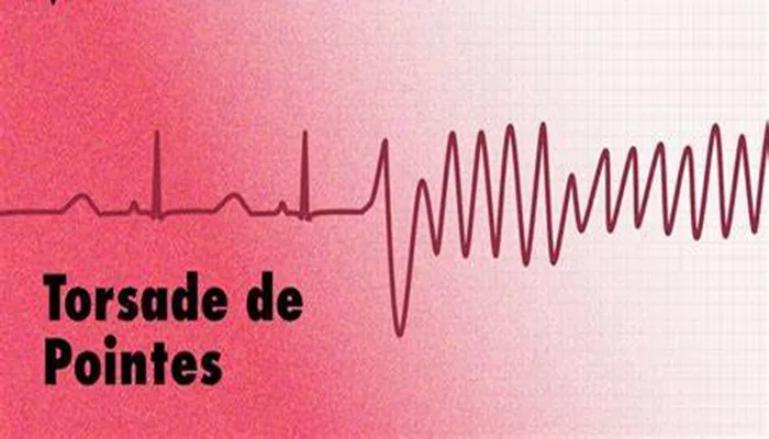
Torsades de Pointes (TdP) is a specific type of polymorphic ventricular tachycardia that is characterized by a unique appearance on the electrocardiogram (ECG) and is often associated with a prolonged QT interval. This arrhythmia can lead to serious complications, including syncope and sudden cardiac death. Understanding the characteristics of torsades de pointes is crucial for early recognition and effective management. This article will delve into the defining features of TdP, its underlying mechanisms, risk factors, clinical presentation, diagnosis, and treatment options.
Introduction to Torsades de Pointes
Torsades de Pointes, which translates from French as “twisting of the points,” was first described by French physician François Dessertenne in 1966. It is a form of ventricular tachycardia that is distinguished by its characteristic ECG findings—specifically, the oscillation of the QRS complexes around the isoelectric line. TdP is typically associated with a prolonged QT interval, which can be congenital or acquired.
The arrhythmia is often self-limiting but can degenerate into ventricular fibrillation, leading to hemodynamic instability and potentially fatal outcomes. Understanding the characteristics of TdP is essential for healthcare providers to manage this arrhythmia effectively and improve patient outcomes.
Characteristics of Torsades de Pointes
1. ECG Findings
The hallmark of torsades de pointes is its distinct electrocardiographic appearance:
Polymorphic QRS Complexes: The QRS complexes in TdP are irregular and vary in amplitude and morphology. They appear to twist around the baseline, creating a visual impression of a “twisting” effect.
QT Interval Prolongation: TdP is associated with a prolonged QT interval, which is the time taken for the heart’s ventricles to depolarize and repolarize. A QT interval longer than 450 ms in men and 460 ms in women is considered prolonged and increases the risk of TdP.
T-Wave Changes: The T waves may also show abnormal morphology, which can precede the onset of TdP.
R-on-T Phenomenon: In some cases, TdP can be triggered by an R-on-T phenomenon, where a premature ventricular contraction occurs during the relative refractory period of the cardiac cycle, leading to TdP.
see also: How Does Long Qt Cause Arrhythmia
2. Clinical Presentation
Patients with torsades de pointes may present with a variety of symptoms, which can range from mild to severe:
Palpitations: Many patients report a sensation of rapid or irregular heartbeats.
Dizziness and Lightheadedness: Due to compromised cardiac output, patients may experience dizziness or lightheadedness.
Syncope: Loss of consciousness may occur, particularly during prolonged episodes of TdP.
Sudden Cardiac Arrest: In severe cases, TdP can lead to ventricular fibrillation and sudden cardiac arrest, necessitating immediate medical intervention.
Mechanisms Underlying Torsades de Pointes
The development of torsades de pointes is primarily linked to the following mechanisms:
1. Prolonged QT Interval
A prolonged QT interval is the most significant risk factor for TdP. The QT interval can be prolonged due to various factors, including:
Congenital Long QT Syndrome: Genetic mutations affecting cardiac ion channels can lead to congenital long QT syndrome, increasing the risk of TdP.
Acquired Long QT Syndrome: Certain medications (e.g., antiarrhythmics, antidepressants), electrolyte imbalances (hypokalemia, hypomagnesemia), and other medical conditions can cause QT prolongation.
2. Early Afterdepolarizations (EADs)
TdP is often triggered by early afterdepolarizations, which are abnormal electrical impulses that occur during the repolarization phase of the cardiac cycle. EADs can lead to triggered activity and initiate TdP, especially in the presence of a prolonged QT interval.
3. Spatial Dispersion of Ventricular Refractoriness
The spatial dispersion of ventricular refractoriness refers to the variation in the refractory periods of different regions of the ventricles.
This can create a substrate for reentrant circuits, which can sustain TdP.
Risk Factors for Torsades de Pointes
Several risk factors can predispose individuals to develop torsades de pointes:
1. Medications
Certain medications are known to prolong the QT interval and increase the risk of TdP. These include:
Antiarrhythmics: Drugs such as sotalol, dofetilide, and quinidine can lead to QT prolongation.
Antidepressants: Some selective serotonin reuptake inhibitors (SSRIs) and tricyclic antidepressants can also prolong the QT interval.
Antibiotics: Macrolides and fluoroquinolones have been associated with QT prolongation.
2. Electrolyte Imbalances
Electrolyte disturbances, particularly low levels of potassium (hypokalemia) and magnesium (hypomagnesemia), can significantly increase the risk of TdP. Maintaining normal electrolyte levels is crucial for cardiac stability.
3. Structural Heart Disease
Patients with underlying structural heart disease, such as heart failure or left ventricular hypertrophy, are at a higher risk for TdP.
4. Genetic Predisposition
Individuals with a family history of congenital long QT syndrome or unexplained sudden cardiac death may have an increased risk of developing TdP.
Diagnosis of Torsades de Pointes
The diagnosis of torsades de pointes is primarily made through electrocardiography:
1. Electrocardiogram (ECG)
An ECG is the gold standard for diagnosing TdP. The characteristic findings include:
Polymorphic Ventricular Tachycardia: The ECG will show the twisting QRS complexes characteristic of TdP.
Prolonged QT Interval: A QT interval greater than 450 ms in men and 460 ms in women is indicative of increased risk.
T-Wave Alternans: Variability in T-wave morphology may also be observed.
2. Patient History and Clinical Assessment
A thorough patient history, including medication use and family history of arrhythmias, is essential for identifying potential risk factors for TdP.
Treatment of Torsades de Pointes
The management of torsades de pointes involves both acute and long-term strategies:
1. Acute Management
In acute cases of TdP, the primary goals are to restore normal rhythm and stabilize the patient:
Magnesium Sulfate: Intravenous magnesium is the first-line treatment for TdP. It helps stabilize the cardiac membrane and suppress early afterdepolarizations.
Electrolyte Correction: Correcting any electrolyte imbalances, particularly hypokalemia and hypomagnesemia, is crucial.
Cardioversion: If the patient is hemodynamically unstable, synchronized cardioversion may be necessary. For pulseless TdP, defibrillation is indicated.
2. Long-Term Management
Long-term management focuses on preventing recurrences and addressing underlying causes:
Medication Review: Discontinuing any QT-prolonging medications is essential.
Beta-Blockers: In patients with congenital long QT syndrome, beta-blockers may be prescribed to reduce the risk of TdP.
Implantable Devices: In high-risk patients, implantation of a cardioverter-defibrillator (ICD) may be considered to prevent sudden cardiac death.
Lifestyle Modifications: Patients should be educated on avoiding triggers, such as certain medications and excessive physical exertion.
Conclusion
Torsades de pointes is a unique and potentially life-threatening arrhythmia characterized by its distinctive ECG findings and association with prolonged QT intervals. Understanding the characteristics of TdP, including its mechanisms, risk factors, clinical presentation, and management strategies, is crucial for healthcare providers. Early recognition and prompt treatment can significantly improve patient outcomes and reduce the risk of complications, including sudden cardiac death.


