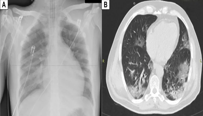
Myocarditis is a condition characterized by the inflammation of the heart muscle, also known as the myocardium. This inflammation can affect the heart’s electrical system and its ability to pump blood, potentially leading to severe consequences such as heart failure, arrhythmias, or even sudden cardiac death. Given the seriousness of myocarditis, early and accurate diagnosis is crucial. However, myocarditis presents a diagnostic challenge because its symptoms can be nonspecific and similar to those of other heart conditions.
One of the questions frequently asked by patients and healthcare professionals alike is whether myocarditis can be detected through a chest X-ray, one of the most common and widely used imaging techniques in medical diagnostics. This article will explore the utility of chest X-rays in diagnosing myocarditis, the limitations of this imaging method, and the role of other diagnostic tools in identifying myocarditis.
Understanding Myocarditis: Causes, Symptoms, And Diagnosis
Myocarditis can result from various causes, including viral infections, autoimmune diseases, and exposure to toxins. Viral infections, particularly those caused by enteroviruses, adenoviruses, and the recent SARS-CoV-2 virus, are the most common causes of myocarditis. Autoimmune diseases, such as lupus or giant cell myocarditis, can also lead to inflammation of the myocardium. Additionally, certain medications, drugs, and environmental toxins can cause toxic myocarditis.
Symptoms of Myocarditis
The symptoms of myocarditis can vary significantly depending on the severity of the condition and the underlying cause.
Common symptoms include chest pain, fatigue, shortness of breath, palpitations, and in severe cases, symptoms of heart failure such as edema (swelling) and fluid retention. However, these symptoms are not unique to myocarditis and can overlap with other cardiac and non-cardiac conditions, making the diagnosis challenging.
SEE ALSO: How Does Clozapine Cause Myocarditis?
Diagnosis of Myocarditis
The diagnosis of myocarditis typically involves a combination of clinical evaluation, laboratory tests, imaging studies, and sometimes invasive procedures such as endomyocardial biopsy. Blood tests may reveal elevated levels of cardiac enzymes, markers of inflammation, and specific antibodies. Imaging studies, including electrocardiography (ECG), echocardiography, and cardiac magnetic resonance imaging (MRI), play a vital role in assessing heart function and identifying signs of myocarditis.
The Role of Chest X-Ray in Diagnosing Myocarditis
What Does a Chest X-Ray Show?
A chest X-ray is a simple, non-invasive imaging technique that provides a visual representation of the structures within the chest, including the heart, lungs, and bones. It is one of the most commonly performed diagnostic tests in clinical practice due to its availability, speed, and relatively low cost. A chest X-ray can reveal various abnormalities such as lung infections, fluid accumulation, tumors, and structural changes in the heart and great vessels.
Can Myocarditis Be Seen on a Chest X-Ray?
While a chest X-ray is valuable in diagnosing many cardiopulmonary conditions, its utility in diagnosing myocarditis is limited. Myocarditis primarily affects the heart muscle at the cellular and tissue levels, and these changes are not typically visible on a standard chest X-ray. Unlike conditions that cause more significant structural changes, such as heart enlargement or pleural effusion, myocarditis does not usually produce distinct or specific findings on an X-ray.
Possible Findings on A Chest X-Ray
In some cases of myocarditis, a chest X-ray may reveal nonspecific findings that suggest the presence of a cardiac condition, but these findings are not definitive for myocarditis. Possible findings on a chest X-ray may include:
Cardiomegaly (Enlarged Heart): Inflammation of the heart muscle can lead to swelling and enlargement of the heart, a condition known as cardiomegaly. While cardiomegaly can be seen on a chest X-ray, it is not exclusive to myocarditis and can occur in various other heart conditions, such as hypertensive heart disease or dilated cardiomyopathy.
Pulmonary Congestion: If myocarditis leads to heart failure, fluid may accumulate in the lungs, resulting in pulmonary congestion or edema. This condition can be visualized on a chest X-ray as increased lung markings, fluid in the lung spaces, or a “batwing” pattern. However, pulmonary congestion is a common finding in heart failure from any cause and does not specifically indicate myocarditis.
Pleural Effusion: In some cases of myocarditis, inflammation may extend to the lining of the lungs (pleura), resulting in pleural effusion, which appears as fluid accumulation in the pleural space on a chest X-ray. Like other findings, pleural effusion is nonspecific and can be caused by various conditions, including infections, heart failure, and malignancies.
Limitations of Chest X-Ray in Diagnosing Myocarditis
Lack of Specificity
The most significant limitation of chest X-rays in diagnosing myocarditis is their lack of specificity. The findings on a chest X-ray may suggest the presence of a cardiac condition, but they do not provide definitive evidence of myocarditis. The absence of specific markers of inflammation or tissue damage on a chest X-ray means that it cannot reliably distinguish myocarditis from other cardiac or pulmonary conditions.
Sensitivity Issues
Chest X-rays also have limited sensitivity in detecting myocarditis, particularly in mild or early-stage cases. Since myocarditis primarily affects the myocardium at the microscopic level, these changes are often too subtle to be detected by a standard chest X-ray. As a result, a normal chest X-ray does not rule out the presence of myocarditis, and further diagnostic testing is usually required.
Dependence on Other Diagnostic Tools
Due to the limitations of chest X-rays, healthcare professionals rely on other diagnostic tools to accurately diagnose myocarditis. These tools include:
Electrocardiography (ECG): ECG can detect abnormalities in the heart’s electrical activity, such as arrhythmias or changes in the ST segment, which may suggest myocarditis.
Echocardiography: This ultrasound-based imaging technique allows for the visualization of the heart’s structure and function. It can detect abnormalities in heart wall motion, ventricular size, and ejection fraction, which may be indicative of myocarditis.
Cardiac MRI: Cardiac MRI is considered the gold standard for non-invasive imaging of myocarditis. It provides detailed images of the heart’s tissue, allowing for the detection of inflammation, scarring, and edema associated with myocarditis.
Endomyocardial Biopsy: In some cases, a biopsy of the heart muscle is performed to confirm the diagnosis of myocarditis.
This invasive procedure involves obtaining a small tissue sample from the myocardium for histopathological examination.
Conclusion
In summary, while a chest X-ray is a valuable tool in the initial evaluation of patients with suspected heart or lung conditions, it is not a reliable method for diagnosing myocarditis. The subtle and microscopic changes associated with myocarditis often go undetected on a chest X-ray, and the findings, if present, are typically nonspecific. As a result, chest X-rays cannot definitively diagnose or rule out myocarditis.
Healthcare professionals must use a combination of clinical evaluation, laboratory tests, and advanced imaging techniques such as cardiac MRI and echocardiography to accurately diagnose myocarditis. These tools provide more detailed and specific information about the condition of the myocardium, allowing for early detection and appropriate management of this potentially life-threatening condition.


