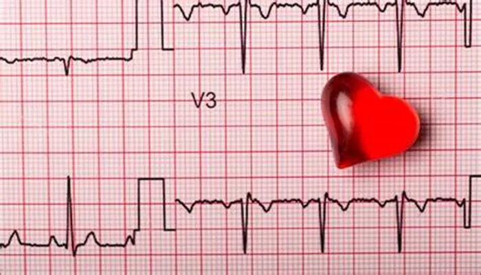
Heart blockage, or heart block, refers to a condition where the electrical signals in the heart are delayed or blocked. This disruption can affect the heart’s ability to beat properly. An electrocardiogram (ECG or EKG) is a crucial tool used by cardiologists to diagnose heart block. This article provides a comprehensive overview of how an ECG detects heart blockage, including types of heart block, ECG findings, and the implications for patient care.
What Is Heart Blockage?
Heart blockage occurs when the electrical impulses that coordinate the heartbeat are interrupted. These impulses travel through pathways in the heart known as the conduction system. The conduction system includes:
The Sinoatrial (SA) Node: Located in the right atrium, it acts as the heart’s natural pacemaker.
The Atrioventricular (AV) Node: Acts as a gatekeeper, slowing the electrical signal before it passes to the ventricles.
The His-Purkinje System: Consists of the His bundle, right and left bundle branches, and Purkinje fibers. It spreads the signal throughout the ventricles.
When these pathways are blocked, the heart’s rhythm can be affected, leading to various forms of heart block.
SEE ALSO: What Is The Survival Rate for Right Coronary Artery Blockage
Types of Heart Block
First-Degree Heart Block: This is the mildest form, where electrical signals are slowed but still reach the ventricles. It is typically detected as a prolonged PR interval on an ECG.
Second-Degree Heart Block: There are two types:
Type I (Wenckebach): The electrical signals progressively slow down until one signal fails to be transmitted to the ventricles.
Type II (Mobitz II): Some signals are blocked, leading to intermittent failure of impulses to reach the ventricles.
Third-Degree Heart Block (Complete Heart Block): None of the electrical signals from the atria reach the ventricles, resulting in a complete dissociation between atrial and ventricular contractions.
How ECG Detects Heart Blockage
An ECG records the electrical activity of the heart over time. It produces a graph with different waves representing various phases of the heartbeat. The key components of the ECG are:
P Wave: Represents atrial depolarization.
QRS Complex: Represents ventricular depolarization.
T Wave: Represents ventricular repolarization.
PR Interval: The time between the start of atrial depolarization and the start of ventricular depolarization.
Diagnosing Different Types of Heart Block Using ECG
First-Degree Heart Block
ECG Findings: The PR interval is prolonged beyond 200 milliseconds (0.20 seconds). This indicates that the electrical signal is taking longer to travel from the atria to the ventricles.
Clinical Implications: Often asymptomatic and may not require treatment. Monitoring may be recommended to ensure it does not progress.
Second-Degree Heart Block
Type I (Wenckebach):
ECG Findings: The PR interval gradually lengthens until a QRS complex is dropped. After a dropped beat, the cycle repeats.
Clinical Implications: May be intermittent and can be asymptomatic. Regular monitoring is important to check for progression to a more severe block.
Type II (Mobitz II):
ECG Findings: The PR interval remains constant before a QRS complex is dropped. The pattern of dropped beats is less predictable.
Clinical Implications: More likely to progress to complete heart block and may require a pacemaker for treatment.
Third-Degree Heart Block (Complete Heart Block)
ECG Findings: No correlation between P waves and QRS complexes.
The atria and ventricles beat independently of each other. The QRS complex may appear wide and abnormal.
Clinical Implications: This is a serious condition that often requires immediate treatment. A pacemaker is typically needed to restore normal heart rhythm.
Additional Considerations in ECG Interpretation
Artifacts: Movement or interference during the ECG can cause misleading results. It is essential to ensure that the ECG is performed correctly and the patient is at rest.
Baseline ECG: Comparing the current ECG with previous records can help identify new or worsening heart block.
Clinical Symptoms: ECG findings should be correlated with symptoms such as dizziness, fainting, or chest pain. Symptoms can provide additional context for the diagnosis.
When to Use An ECG
An ECG is often used in various clinical scenarios to assess heart function and diagnose potential problems, including:
Routine Check-ups: To monitor heart health, especially in patients with risk factors or symptoms suggestive of heart disease.
Preoperative Assessments: To evaluate the heart’s electrical activity before undergoing surgery.
Emergency Situations: To quickly diagnose heart block and other cardiac conditions in patients presenting with chest pain, palpitations, or syncope.
Treatment Options
Treatment for heart block depends on its severity and the presence of symptoms:
First-Degree Heart Block: Usually does not require treatment but should be monitored regularly.
Second-Degree Heart Block: Type I may require less intervention, while Type II often necessitates a pacemaker to prevent progression to complete heart block.
Third-Degree Heart Block: Requires immediate intervention with a pacemaker to regulate the heart’s rhythm and ensure adequate blood flow.
Conclusion
An electrocardiogram is a vital diagnostic tool for detecting heart blockage. By analyzing the electrical activity of the heart, an ECG can identify various types of heart block and guide appropriate treatment. Understanding the types of heart block and their ECG characteristics is essential for effective diagnosis and management, ensuring better outcomes for patients with this condition.

