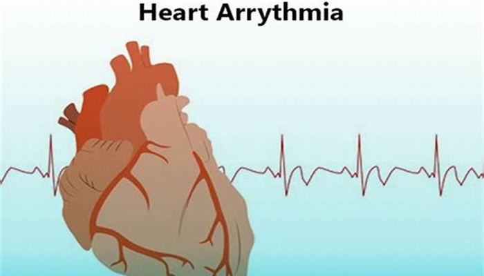
Arrhythmias frequently arise after radiotherapy for nonsmall cell lung cancer, but recent advances in treatment have led to notable increases in median survival rates.
A collaboration of doctors from Cedars-Sinai Medical Center in Los Angeles and Brigham and Women’s Hospital in Boston, looked to analyze the association of distinct arrhythmia classes with cardiac substructure radiotherapy dose in patients with locally advanced nonsmall cell lung cancer.
The study, which was published in JACC: CardioOncology on Aug. 6, 2024, was led by Katelyn M. Atkins, M.D., radiation oncologist at Cedars-Sinai Medical Center and identified specific dose-volume predictors for different types of arrhythmias.
Raymond H. Mak, M.D., director of patient safety and quality director of clinical innovation at Brigham and Women’s Hospital and an associate professor at Harvard Medical School, who was part of the research team, explained that over the last eight years, the research group has been elucidating the connection between radiation exposure to the heart as part of lung cancer treatments, and the effects on these patients in terms of the risks for major cardiac complications.
“Historically, before we started this work, a lot of people thought lung cancer prognosis was so terrible, it was such a difficult to treat cancer that didn’t matter what you did to these patients,” he said. “Cardiac health complications wouldn’t occur until five to 10 years after the cancer treatments completed.”
However, the work of this collaboration of oncologists has demonstrated that cardiac complications are common in these patients.
“This paper builds on those prior discoveries focusing on one specific kind of complication of cardiac arrhythmias—either a rapid heartbeat or abnormal heartbeat,” Mak said. “What we’ve done that is different is we’ve mapped radiation exposures to different parts of the heart to predict the risk for each of the different kinds of arrhythmias.”
The team conducted a retrospective analysis of 748 patients with locally advanced nonsmall cell lung cancer treated with radiotherapy and cardiac substructure dose parameters were calculated.
“We used artificial intelligence (AI) tools to help us identify and measure the dose radiation exposure into different parts of the heart, like the pulmonary veins and parts of the heart that are difficult to draw,” Mak said. “The use of AI accelerated our research; something that may have taken a year or two to identify the connections, literally took weeks.”
Mak explained that receiver-operating characteristic curve analyses for predictors of common terminology criteria for adverse events grade 3 or greater atrial fibrillation (AF), atrial flutter, non-AF and non–atrial flutter supraventricular tachyarrhythmia (SVT), bradyarrhythmia, and ventricular tachyarrhythmia (VT) or asystole were calculated.
The data revealed that 17.1% of patients experienced grade 3 or greater arrhythmias. Furthermore, the researchers found pathophysiologically distinct arrhythmia classes associated with radiotherapy dose to discrete cardiac substructures, including PV dose with AF and SVT, left circumflex coronary artery dose with atrial flutter, right coronary artery dose with bradyarrhythmia, and left main coronary artery dose with VT or asystole, guiding potential risk mitigation approaches.
“Now when we design a radiation treatment for a lung cancer patient, we can now understand that the pulmonary veins, as an example, is an important risk predictor, and can design radiation plans to avoid giving excess radiation dose to those pulmonary veins to reduce the risk of the presentation developing atrial fibrillation after the radiation treatment,” Mak said.

